Frequency of Seven Common Deletion Alfa-Globins Mutation Carriers in Suspect Referred In West Mazandaran, Iran
Zahra Nashtahosseini, Ali Nazemi, Shahrbanoo Keihanian, Reza Hajihosseini, Mohammad Eskandari
| Zahra Nashtahosseini1,*, Ali Nazemi2, Shahrbanoo Keihanian3, Reza Hajihosseini4, Mohammad Eskandari5 1 MSc in Biochemistry, Payame Noor University (PNU).Tehran, Iran; 2 PhD in Molecular Genetics, Assistant Professor, Department of Biology, Islamic Azad; University–Tonekabon branch, Mazandaran, Iran; 3 Department of Oncology,Islamic Azad University -Tonekabon branch, Mazandaran, Iran; 4 PhD in Biochemistry, Professor, department of biology, Payame Noor University (PNU).Tehran, Iran; 5 MSc in Genetics, Department of Biology, Gilan University, Iran. |
| Corresponding author. Tel: +98-09112928720; E-mail: z_nashtahoseini@yahoo.com |
| Received: December 22, 2015 Accepted: January 07, 2016 Published: January 14, 2016 |
| Citation: Nashtahosseini Z, Nazemi A, Keihanian S, et al., Frequency of Seven Common Deletion Alfa-Globins Mutation Carriers in Suspect Referred In West Mazandaran, Iran. Electronic J Biol, 12:1 |
Abstract
Frequency of mutation in alpha-globins gene has a great diversity in different populations, and unlike other hemoglobinopathies is not recognizable by simple biochemical experiments. Carriers of these genes have anemia microcytic and its clinical syndromes are variable from being without any sign to fatal hemolytic anemia. Diagnosis of these carriers is useful in order to find out what causes microcytic, avoid long-term treatment of iron deficiency anemia, and prevent marriage between transporters of alpha thalassemia and birth of infants suffering from severe anemia.
In this study, 136 blood samples of people with reduced blood indexes HBA2 Á MCH<27pg Á MCV<80fl were gathered and studied randomly from laboratories in Tonekabon, Ramsar, and Chalous. Identification of deletion mutations is done by sage of multiplex- PCR method. Deletion mutations include single gene deletions -α3.7 and -α4.2, homozygous double gene deletions -α3.7/-α3.7 and - -THAI in 56 samples of all studied samples. Also, a triple gene deletion -α3.7/- - FIL, which has never been recognized, is identified in this study. The most prevalent deletion in Tonekabon is single gene deletion -α3.7. This fast, low-cost, and simple method is useful for status determination of those who are indistinctively classified in globin alpha gene.
Keywords |
||||
| Alpha thalassemia; Multiplex PCR; West mazandaran. | ||||
Introduction |
||||
| Heterogeneous group thalassemia syndromes are heredity anemia, which are the results of defection in structure of one or multi globin chain [1]. Alpha thalassemia is a beaten autosomal disorder and the most prevalent synthesis hemoglobin heredity disorder in the world [2]. Globin alpha category is on 16th chromosome and α1 and α2 are its main genes, which their nucleotide sequences are very similar [3]. The rate of disorder in globin alpha chain`s synthesis under the effect of mutation depends on number of deactivated genes and involvement of α1 and α2 genes [4]. Deletion mutations on globin alpha genes result in deletion of either both alpha genes α° or just one gene α+. There are five prevalent deletion mutations with creating α° thalassemia, which are: -- FIL, -- MED, -- THAI, -- SEA, --20.5. The most prevalent of them are -- SEA in south-east of Asia, -- MED in Mediterranean, and -- THAI in Taiwan with a widespread prevalent. Recently, the -- FIL mutation type has been seeing in those countries. Gene mutations are created by deletion of a alpha globin gene as α+. There are two α+ gene mutations: -α3.7, - α4.2. | ||||
| Based on the construction rate of globin alpha chain, alpha thalassemia is divided into four clinical groups: the first group is the state of silent carrier α+, which is deactivated in only one gene of four alpha genes in the patient, and in this state, the globin alpha chain reduces a little and these people are hematologically normal. | ||||
| The second group is the state of alpha thalassemia trait α°, which is created by the deletion of two active alpha globin genes. These people suffer from slight hypochromic and microcytic anemia. The third group is people with hemoglobin H disease, which is created because of α+ and α° heterozygotes and create just one alpha chain. Those people suffer from sever hemolytic anemia, jaundice, Splenomegaly [5]. In the fourth group, hydropas fetalis state, no alpha chain is created. In this state, embryo`s hemoglobin is a tetramer from gama chain which is called hemoglobin barts [4]. | ||||
| In this study, molecular identification of seven prevalent deletion mutation alpha globin gene cluster is done in people who were suspect to alpha thalassemia with HbA2ÁMCH<27fl) (MCV<80 pg reduced natural blood indexes. | ||||
2. Materials and Method |
||||
| 136 blood samples with MCV and MCH lower than normal level and HbA2 lower than 3.5%, who had been going to diagnostic laboratories in Tonekabon, Chalous, and Ramsar during one year (1389), is gathered. Genomic DNA is extracted from 0.5 ml whole blood that was gathered in tubes of Anticoagulants EDTA by DNA extraction kit (Roche). Then, multiplex - PCR reaction performed on samples based on Tan method [6] in order to identify prevalent deletion mutations. The reaction in the volume of 25 micro litter which contains (HCl, 20 mM KCl, 5 mM (NH4)2SO4, 2 Thermal proliferation cycles in the PCR device of BioRad Company was done in the form of primary denaturation for the length of 5 minutes in 95°C and was followed by 35 cycles, 95°C for the length of 1 minute, 60°C for the length of 1:30 minute, 72°C for the length of 2:15 minutes, and final expansion in the temperature of 72°C for the length of 5 minutes [6]. After the end of reaction, 10 micro litters of PCR products was electrophoresed on the one percent agarose gel in the TBE buffer with the marker of 100 to 3000 pair play. Then the gel was colored with ethidium bromide color. | ||||
3. Findings |
||||
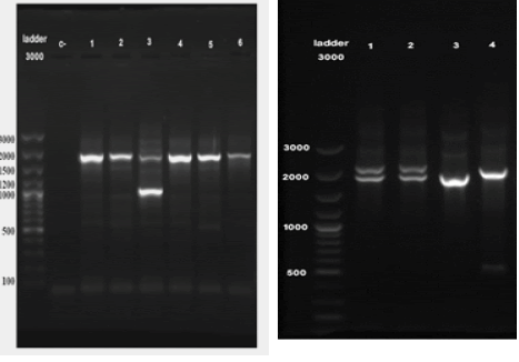
| Identifying of the most prevalent known deletions of alpha globin gene are getting done through proliferation of areas which include alpha globin gene mutation through multiplex- PCR method and study on agarose gel and observation on presence or absence of specific positions. This method provides the possibility of identifying known globin alpha deletions at the same time and in a reaction tube. In this study, identifying of seven prevalent alpha thalassemia deletion (--FIL, -α20.5, -- MED, -- SEA, -- THAI, -α4.2, -α3.7) was done on 136 samples with lower than normal MCV and MCH and level of HbA2 lower than 3.5%. From all studied samples, in 56 samples deletion mutation was identified and in 80 samples none of these deletion mutations was observed. Type and distribution of elimination mM Tris-), 0.2 millimolar mixed dNTP, 1.5 millimolar magnesium chloride 50 millimolar, 5 micro litter Q solution (Q-Solution, Qiagen), 2 unite of Hot start taq DNA Polymerase enzyme, and seven primer pairs with final densities that are mentioned in the Table 1.mutations in this area is shown in Table 2. In the study of 7 prevalent deletions in alpha globin cluster with Multiplex-PCR method in Tonekabon area, the most prevalent were -α3.7 with 36% and then -α4.2 with 3.67% (Table 2) (Figure 1). | ||||
4. Conclusion |
||||
| The rate of prevalence of thalassemia syndromes is 4.8% in the world. The study of scattering of this disease show that in addition of Mediterranean countries, which in thalassemia was diagnosed for the first time, this disease is seen in Asia and the Middle East [7]. Continuing immigration of people, now days, there is no country in the world which this disease has not been affected some percent of its people. In Iran, especially northern Iran, because of high prevalence of thalassemia, the need of identifying different mutations and their frequencies is essential [8]. This study shows more than 41.15% of known alpha thalassemia cases are caused by deletion of one or double gene of alpha globin from 16th chromosome. Probably, 58.82% of samples have non-deletion mutations or deletion mutations other than these seven prevalent deletions. As a result, more study on healthy samples suspect to alpha thalassemia is necessary. | ||||
| This study shows mutation of -α3.7 in 44 (36%) studied samples represents the highest frequency of deletion mutation in Tonekabon area. Mutation of -α4.2 is diagnosed in 5 (3.67%) samples. This is the second prevalent deletion mutation in Tonekabon area. In addition, --THAI/αα double deletion is identified in this area in addition of one elimination of α3.7/--FIL, which has not been identified so far. This person with this genotype has triple deletion in alpha globin gene and is suffering from hemoglobin H disease. | ||||
| In the studies of other researchers in other parts of the country, frequency of -α3.7 mutation is reported as 32.8%, 42.5%, and 44.9% between suspect people [9-11]. Also, mutation frequency of -α4.2 in the civilization study was reported as 9.1% and the second type of prevalent mutation was diagnosed (9). Although Neishaboor7i and associates reported the mutation frequency of -α3.7 (31.6%), no -α4.2 mutation was reported [12]. | ||||
| Multiple- PCR method, which is innovated by Tan and associates in 2001, is a low cost, fast, available, and without radioactive method. Having special features, this method is useful for molecular determining and describing and prevention of repeating high-cost analysis or long-term iron treatment in population screening. There should be more studies on those samples which were lacking these mutations and Real time- PCR or Sequencing DNA can be used for this purpose. Identifying people with alpha thalassemia mutations is very important in order to prevention of giving birth to children with hemoglobin H and hydrops fetalis disease in addition of social, economic, mental, and sanitary issues that these children`s families and society will be facing. In addition, those mothers who are having embryos with hydrops fetalis, are at 80% risk of getting high blood pressure in pregnancy period, bleeding and preterm birth, depression during pregnancy, difficult childbirth, retained placenta, and bleeding after childbirth. Mentally, carrying a dead embryo till the time of birth is very difficult for these mothers [13]. | ||||
| Being simple and comprehensive, this method is significantly low cost and complexity of screening for prevalent alpha thalassemia deletion is reduced [14]. Considering high prevalence of thalassemia disease, this new information is useful for effective and available prevalent alpha thalassemia deletion screening in Tonekabon area. | ||||
5. Appreciation |
||||
| This study was conducted in the research lab of Islamic Azad University in Tonekabon. We thank everyone who has helped us conducting this project and gathering samples. | ||||
Tables at a glance |
||||
|
||||
Figures at a glance |
||||
|
||||
References |
||||
|

Open Access Journals
- Aquaculture & Veterinary Science
- Chemistry & Chemical Sciences
- Clinical Sciences
- Engineering
- General Science
- Genetics & Molecular Biology
- Health Care & Nursing
- Immunology & Microbiology
- Materials Science
- Mathematics & Physics
- Medical Sciences
- Neurology & Psychiatry
- Oncology & Cancer Science
- Pharmaceutical Sciences

