Organotypic Brain Slice Triple Culture of Neocortex-striatum-substa-nigra of Neonatal Rats
Jiao Shi, Weijun Yu, Guiyuan Sun, Suwen Zhu
1Department of Histology and Embryology,China Medical University,Shenyang,Liaoning 110001,China
2Department of Animal Science,Shenyang Agriculture University,Shenyang,Liaoning 110161,China
Abstract
To investigate in vitro organotypic brain slice triple culture of neocortex-striatum-substantia nigra of postnatal rats. Slices (300um) from 2-day-old Wistar rats were transferred into an incubator with insert of Millicell membrane and incubated in Hanks balanced solution. They were cultured for 0d, 10d, 20d and 30d and were observed under a converted microscope to evaluate the growth state. Dopamine neurons and axons were labeled by tyrosine hydroxylase (TH) immunofluorescence. The ability that the slices absorb EB was tested with LCM510. Axons of TH-positive neurons in the substantia nigra were grown into the striatum. On 20d, the striatum join together with the substantia nigra with no necrotic cells found. On 30d, 10 percent neurons died of EB and necrosis. It was demonstrated in our experiment that 20d triple cultures with normal pathway formed from the substantia nigra to the striatum and this provides a unique in vitro histological model for studying the mechanism of some neural retrograde diseases such as Parkinson disease.
Keywords
Brain slice triple culture,Disease model,Parkinson disease
1. Introduction
To study nervous system disease,one must establish a disease model,which include stiring liaison net between inherent neuron of vitality. For example,according to pathological feature of parkinson disease’s (PD),different models have been established,such as traditional 6-OHDA model and MPTP model [1-3]. All these models can qualitatively reproduce changes of pathology and behaviour of PD,but have a long periodicity and low success (only 40%). At the same time,most difficulties in vivo involved a great deal of uncontrlled factors and experimentation condition. Despite of the above mentioned difficulties,we want to establish a good model of organotypic cortex- striatum-mesencephaln in vitro. Our research is described in detail as below.
2. Materials and methods
2.1 Brain slices triple culture
For the preparation of the triple cultures,coronal sections (300μm) from rat brains at postnatai day 0-2 (Wistar) were cut on a vibratome. Slices containing neostriatum,stratum and cortex,cultured on a Millicell-CM membrane,were randomly divided into three groups according to 10d,20d and 30d.First Millicell-CM membranes were layed in 6 well cell culture cluster,which including 1ml manpower liquid of brain (mmol/L: NaCL126,KCL 2.5,NaH2PO3 1.2,MgCL 1.3,glucose 11,NaCO3 25,CaCL2.4).Then put cortex,stratum and neostriatum slices in turn. Finally aspirate manpower liquid of brain and affiliate culture medium (85 percent high glucose DMEM,15 percent standard moggy serum).The Culture medium just reach horizontal of slices and didn’t overflow. Slices were observed by inverted microscope and pictured at 10d,20d and 30d.
2.2 LCM510 focused on vigor of slides
EB decoration method: EB can dye dying cell and active cell cannot be dyed,so we put the 10d、20d and 30d brain slice in medium containing 1μlEB (density of 100μg/ml) for30min. EB dye dying cell red,which were fault scanning with LCM510. We count died cell (%)within a radius of 100μm and an optical thickness.
2.3 Immunohistochemistry
SABC method was performed. For immuno-histochemistry and neurobiotin reconstruction,trple cultures were fixed 4% paraformaldehyde overnight at 4℃,and then incubated in 2% H2O2 in phosphate buffered saline (PBS; 5min; 3times) and 0.3% Triton-x (37℃; 1hour). The underlying membrane with cultures was mounted on slides. The cultures were incubated overnight with a mouse monclonal antibody against TH (Incstar; 1:500) in PBS. The cultures were then incubated in Texas red anti-mouse IgG (4℃,12hour) and observed with a fluoresc-microscope (Motic; 400). At the same time slices of 10d,20d and 30d were took occasionally and fixed (0.25%glutaraldenhyde),Ultrathin section of brain fixed slices were scanned through electron microscope
3. Results
3.1 Results in inverted microscope
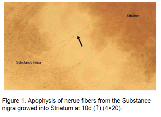
Slices cultured of 10d got thinner with growth coronal. The fibres of neuron from the stratum growth to neocortex. All this demonstrated the brain slices were brimming with vigor. (Figure 1). When 20d,striatum and substance nigra were grown single cultures. In 30-day,cultured slices stopped growth and glial proliferation were found.
3.2 results of TH immunohistochemistry EB decoration
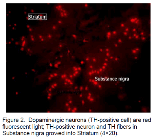
TH-positive cells were characterized by their large fusiform or polygonal cell bodies and 2-5 primary dendrites that eventually branch into higher order dendrites. In 20d a TH positive fiber network was found in the neostriatum. The fibres of TH-positive neuron and TH-positive neuron from the substa-nigra growth to striatum (Figure 2 ).
Figure 2 . Dopaminergic neurons (TH-positive cell) are red fluorescent light; TH-positive neuron and TH fibers in Substance nigra growed into Striatum (4×20).
Results of EB decoration: At 10d and 20d,brain slices weren’t dyed by EB there was no red fluores-cell in brain slices.But at 30d,about 10percent cell died and were dyed red by EB.
3.3 results of Ultrathin section of brain slices
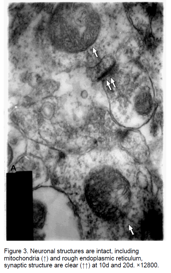
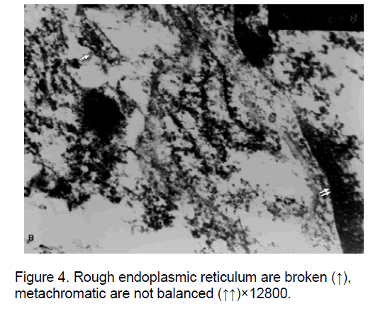
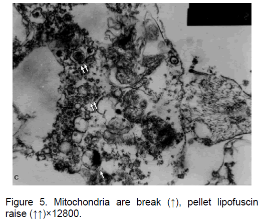
Ultrathin section of brain slices were scaned through electron microscope.Neuronal structures are intact,including mitochondria and rough endoplasmic reticulum,synaptic structure are clear at 10d and 20d (Figure 3 ); Rough endoplasmic reticulum are broken,metachromatic are not balanced,mitochondria are broken,pellet lipofuscin raise at 30d (Figure 4 and Figure 5 ).
4. Discussion
The gateway of the stratum and neostriatum is in correspondance with Parkinson disease,which pathological change located in the gateway stratum and neostriatum [3]. Brain slice triple culture of neocortex-striatum-substa-nigra on Millicell-CM membrane,are found to be the correlational slice in vitro. In theory,Triple brain slice worked on each other in cultured environment,keeping integrated touching loop,recieving distribution and nerve function. A part of the brain slice is immerged in a cultured medium,while the remainding part is exposed to air. This was propitious to brain slice ingesting nutrition and oxygen from medium and air. We can see from the results that nerve cell in brain slice is active at 10d and 20d. The fibres of TH-positive neuron and TH-positive neuron from the stratum grow to neocortex. By inverted microscope scanning,substa-nigra neuron axon together with striatum,and combining with TH Immunohistochemistry dyeing,the results suggest that neuron axon above was TH positive fiber. At the same time,TH-positive neurons were found in striatum. So we concluded that brain slice culture model may use for pathogenesis and medication of PD.
Connelly et al. adopted MTT method to quantitatively analyze the living tissure [4]. Owing to the thickness of brain slice,it is difficult to count living cell with MTT method. We check brain slice vigor with EB decoration method. EB can dye a dying cell and but not the living cell,Currently 1:l0000 EB can be exposed to visible light .so when dying cell was decorated by EB,we can scan with LCM510. So this method is novel and simple.
In cultured brain slice without blood-brain barrier,drug immediately acts on brain tissue on independence of solid location. Furthermore we can change circumstance by intervention of drug. It is known that individual brain slice could repeat natural electricity physiology of an animal body[5].So,culture method of brain slice and spinal cord slice were found in many domestic or overseas laboratory [6,7].
We conclude that the method of the brain slice triple culture of neocortex-striatum-substa-nigra on Millicell-CM membrane is a simple and effective nerve tissue culture method in vitro. This method may be used for PD investigation.
References
- Sauer H.,Oertel W.H. (1994) Progressive degeneration of nigrostriatal dopamine neurons following intrastriatal tertminal lesions with 6-hydroxydopamine a combined tracing and immunocytochemical study in the rat. Neuroscience,59: 401-410.
- Betarbet R.,Sherer T.B.,Greenamyre J.T. (2002) Animal models of Parkinsons disease. Bioessays,24: 308-318.
- Betarbet R.,et al. (2000) Chronic systemic pesticide exposure reproduces features of Parkinson’s disease. Neuroscience,3: 1301-1310.
- Connely C.A.,Chen E.,Colquhoun S.D. (1999) Metabolic activity of cultured rat braimtem,hippocampal and spinal cord sliees. Neurosci Mahods,99(1-2): 1-7.
- Stoppini L.,Buchs P.A.,Muller D.A. (1991) Simple method for organotypic cultures of nervous tissue. Neurosci Methods,37(2):173-180.
- laake J.H.,Haug F.M.,Wieloeh T.,et al. (1999) A simple in vitro model of ischemic based on hippocarnpal slice cultures and propidum iodide fluorescence. Brain Res Protos,4(2): 173-184.
- Morrison B.,Eberwine J.H.,Meaney D.F.,et al. (2000) Traumatic injury induces differential expression of cell death genes in organotypic brain slice cultures determined by complementary DNA array hybridization. Neuroscience,96(1):131-140.

Open Access Journals
- Aquaculture & Veterinary Science
- Chemistry & Chemical Sciences
- Clinical Sciences
- Engineering
- General Science
- Genetics & Molecular Biology
- Health Care & Nursing
- Immunology & Microbiology
- Materials Science
- Mathematics & Physics
- Medical Sciences
- Neurology & Psychiatry
- Oncology & Cancer Science
- Pharmaceutical Sciences
