Evaluation of the Clinical Significance of Transforming Growth Factor-ÃÆà ½Ãâââ¬â¢ and Vascular Endothelial Growth Factor Expressions in the Synovial Tissue of Patients with Osteoarthritis
Ding Hao
Department of Orthopaedics, The First Affiliated Hospital, School of Medicine, Shihezi University, China.
- *Corresponding Author:
- Tel: 13260380656
Email: 1253058993@qq.com
Received date: March 25, 2017; Accepted date: March 29, 2017; Published date: April 05, 2017
Citation: Hao D. Evaluation of the Clinical Significance of Transforming Growth Factor-Β and Vascular Endothelial Growth Factor Expressions in the Synovial Tissue of Patients with Osteoarthritis. Electronic J Biol, 13:1
Abstract
Background: Knee osteoarthritis is a common chronic degenerative disease resulting in joint histopathologic and serologic changes. TGF-β and VEGF change have a close relationship with knee osteoarthritis. Objective: To evaluate the expression of Transforming Growth Factor (TGF)-β and Vascular Endothelial Growth Factor (VEGF) in patients with and without knee Osteoarthritis (OA) and to assess the clinical relevance between these expressions and knee OA. Design: Case-control study. Setting: Hospital. Methods: We collected pathological knee synovial tissue from 50 patients with OA. We evaluated TGF-β and VEGF expressions of synovial tissue using immunohistochemical methods. The χ2 test was used for statistical analysis. Participants: Patients with and without knee OA were compared. We divided patients with OA into mild, moderate, and severe groups (n=50 per group). The control group included 50 patients without arthritis. Results: We found inflammatory infiltrates, synovial cell hyperplasia, significant blood capillary hyperplasia, fiber hyperplasia around the capillaries, and disordered arrangement of cartilage cells in the synovial tissue of patients with OA. TGF-β expression increased in patients with the degree of knee synovial tissue with OA; the increased VEGF expression correlated with blood capillary hyperplasia and inflammatory infiltrates. TGF-β and VEGF expressions were significantly different between the control and severe groups (P<0.01). There was no obvious difference between the control and mild groups, mild and moderate groups, and moderate and severe groups. TGF-β and VEGF expressions had a significant positive correlation (r=0.783; P<0.05). Conclusion: TGF-β and VEGF expressions are related to the degree of synovial lesions of knee joints with OA; thus, they can be regarded as an indicator of knee OA.
Keywords
Knee joint; Osteoarthritis; Synovial tissue; Transforming growth factor beta; Vascular endothelial growth factors.
1. Introduction
Knee osteoarthritis (OA) causes articular cartilage damage and it is a type of degenerative disease and inflammatory synovial joint disease characterized by the destruction of articular cartilage, articular surface osteophyte formation, synovial cell hyperplasia, synovial inflammation and joint gap narrowing [1]. OA is one of the most common forms of arthritis, and it is the main cause of musculoskeletal disease worldwide [2].
Vascular Endothelial Growth Factor (VEGF) is one of the most studied cell factors, and its functions include the regulation of blood vessels and angiogenesis [3]. According to previous research studies, VEGF can cause increased vascular permeability, a series of inflammatory phenomena, endothelial cell hyperplasia, increased expression of inter-cell molecular adhesion, and inhibition of endothelial cell apoptosis, among others [4,5]. In addition, VEGF can promote an animal’s neuron axon regeneration in the brain cortex and nerve cell protection through interaction with specific receptors. The Transforming Growth Factor (TGF) is a conversion factor, and functions in cell hyperplasia, differentiation, apoptosis, mobility, adhesion, extracellular matrix formation, wound healing, and bone reconstruction. TGF-β performs its biological functions through interaction with receptors.
The causes of deterioration in OA are multifactorial. To better understand the occurrence of synovial tissue lesions and their development in OA patients and to provide guidance for the clinical treatment of OA, we focused on the relevant factors in knee joint synovial lesions in OA patients. Our goal was to find an efficient way to treat OA through the detection of knee joint synovial levels of TGF-β and VEGF.
The present study evaluated TGF-β and VEGF expressions in patients with and without knee OA and assessed the relevance between these expressions and knee OA of synovial tissue.
2. Materials and Methods
Patients from January 2010 to December 2014 diagnosed with knee OA were treated with arthroscopic surgery at our hospital, and they were divided into mild, moderate, and severe groups according to the severity of knee OA (n=50 per each group; total, 150 patients; age range, 32-50 years; 102 men and 48 women; Table 1). Patients without knee lesions on arthroscopy were included in the control group (total, 50 patients; age range, 25-50 years; 26 men and 24 women).
We categorized the disease period into early, middle and late phases and used the Outerbridge classification of articular cartilage damage under arthroscopy [6]. We considered grade I as early and mild lesions group, grades II-III as middle and moderate lesion group, and grade IV as late and severe lesion group.
2.1 Inclusion and exclusion criteria
Consecutive patients were prospectively enrolled if they were adults undergoing arthroscopy of the knee with a presumptive diagnosis of osteoarthritis. Patients were excluded if they had concomitant liver or kidney dysfunction, cancer, or infectious disease, or had laboratory testing showing positive rheumatoid factor, elevated sedimentation rate, or elevated C-reactive protein.
2.2 Main chemical reagents
Tris-buffer saline, diaminobenzidine chromogenic agent, tris-hydrochloride, wood stain, acid alcohol, microwave antigen-repairment liquid, eosin stain and immunohistochemical reagents such as xylene were used to assess TGF-β and VEGF expressions of patients’ knee joint synovial tissue (Table 2).
2.3 Immunohistochemical antibodies
TGF-β and VEGF antibodies were purchased from Bioss Antibodies, Inc. (Woburn, Massachusetts, USA) (Table 3).
2.4 Experimental equipment
The following experimental equipment was used: an arthroscopic system (Stryker 597T, Stryker Corporation; Kalamazoo, Michigan, USA), constant temperature drying oven (GW202V, Shanghai Experimental Instrument Company, Shanghai, China), microscope (CX31 RBSF; Olympus, Tokyo, Japan), fast vortex mixer (XK96-A,Shanghai Bilang Instrument Company, Shanghai, China), trace oscillator (75-2A, Shanghai Medical Analysis Instrument Factory, Shanghai, China), and paraffin section machine (2135, Leica, Boston, MA).
2.5 Evaluation of synovial tissue
The synovial tissue obtained arthroscopically was sliced for hematoxylin and eosin staining. We observed the pathological and physiological changes of the joints, and evaluated TGF-β and VEGF expressions, using the immunohistochemical method; the operation was performed as described previously. The absorbance value was measured at a 450 nm point, using a standard enzyme instrument, and we determined the results according to the standard concentration and absorbance value. Finally, we calculated TGF-β and VEGF expression levels according to the absorbance value of each sample.
| Classification | Characteristics under Arthroscopy |
|---|---|
| I | Soft and swelling |
| II | Broken and cracked bone is less than 1.3 cm in diameter |
| III | Broken and cracked bone is greater than 1.3 cm in diameter |
| IV | The subchondral bone is exposed |
Table 1. Articular cartilage injuries according the Outerbridge classification [6] (arthroscopic observation).
| Positive Percentage (%) | Score | Staining Strength | Score | Total | Result |
|---|---|---|---|---|---|
| 0-10 | 0 | None | 0 | 0 points | Negative |
| 10-40 | 1 | Weak | 1 | 1-2 points | Weak positive |
| 40-70 | 2 | Moderate | 2 | 3-4 points | Positive |
Table 2. Standard grading of TGF-β and VEGF expressions.
| Antibody Name | Concentration | Source of the Product |
|---|---|---|
| TGF-β | 1:600 | Bioss Antibodies, Inc. |
| VEGF | 1:200 | Bioss Antibodies, Inc. |
Table 3. Antibody name, origin, and concentration.
2.6 Ethical consideration
The Regional Ethics Review Board in Shihezi, China approved the study. All participants received written and verbal information about the study before giving their written consent to participate in accordance with The Code of Ethics of the World Medical Association.
2.7 Statistical analysis
Using SPSS version 13.0 statistical software (IBM Corp., Armonk, NY, USA), group differences were analyzed using the χ2 test (α=0.01). Multiple comparisons among the groups were assessed using the χ2 segmentation method (α=0.01). A correlation between TGF-β and VEGF expressions was determined if there was a linear trend between the two variables (α=0.01).
3. Results
3.1 Histological chemistry analysis
In the control group, the synovial layer consisted of 1-3 layers of synovial cells, and fewer synovial cells were found in the lower layer. In the experimental group, synovial layer thickening, 3-10 layers, significant blood capillary hyperplasia, inflammatory cell infiltration, and hyperplasia of fibrous tissue were observed.
3.2 Immunohistochemical observations
VEGF expression
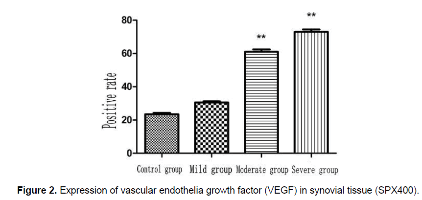
The positive expression of VEGF was indicated as tan particles and these particles were distributed in vascular endothelial cells, synovial epithelial cells, and stromal cells. We analyzed VEGF-related data using the chi-square test, and the positive result was significantly different among the study groups (P=0.01). Synovial pathological changes correlated with the expression of VEGF (χ2 =12.516, P=0.007). We analyzed each difference using the chi-square test, and we found that the positive rate of VEGF expression was significantly higher in the groups with synovial pathological changes (P<0.01; Figures 1 and 2).
| Group | Number of Specimens | VEGF (Positive) | TGF-β (Positive) |
|---|---|---|---|
| Control group | 50 | 7 (14%) | 12 (24%) |
| Mild group | 50 | 15 (30%) | 15 (30%) |
| Moderate group | 50 | 32 (64%) | 30 (60%) |
| Severe group | 50 | 40 (80%) | 38 (76%) |
*P<0.01.
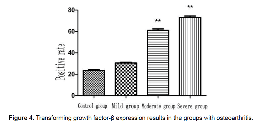
Table 4. Vascular endothelial growth factor (VEGF) and transforming growth factor (TGF)-β expression test results of each group of (%).
Figure 1. Expression of vascular endothelia growth factor (VEGF) in synovial tissue (SPX400). A: Negative expression of VEGF in synovial tissue; B: Weak positive expression of VEGF in synovial tissue; C: Positive expression of VEGF in synovial tissue; and D: Strong positive expression of VEGF in synovial tissue.
TGF-β expression of each group
The positive expression of TGF-β material was indicated as tan particles, and they were distributed in vascular endothelial cells, synovial epithelial cells, and stromal cells. We analyzed TGF-β-related data using the chi-square test, and its positive result was significantly different among the groups (P=0.01; Table 4). Synovial pathological changes correlated with the expression of TGF-β (χ2 =17.817, P=0.003; Table 5). We analyzed each difference using the chisquare test and we found that the positive expression of TGF-β was significantly higher in the groups according to the degree of synovial pathological changes (P < 0.01; Figures 3 and 4).
3.3 Correlation between VEGF and TGF-β expressions
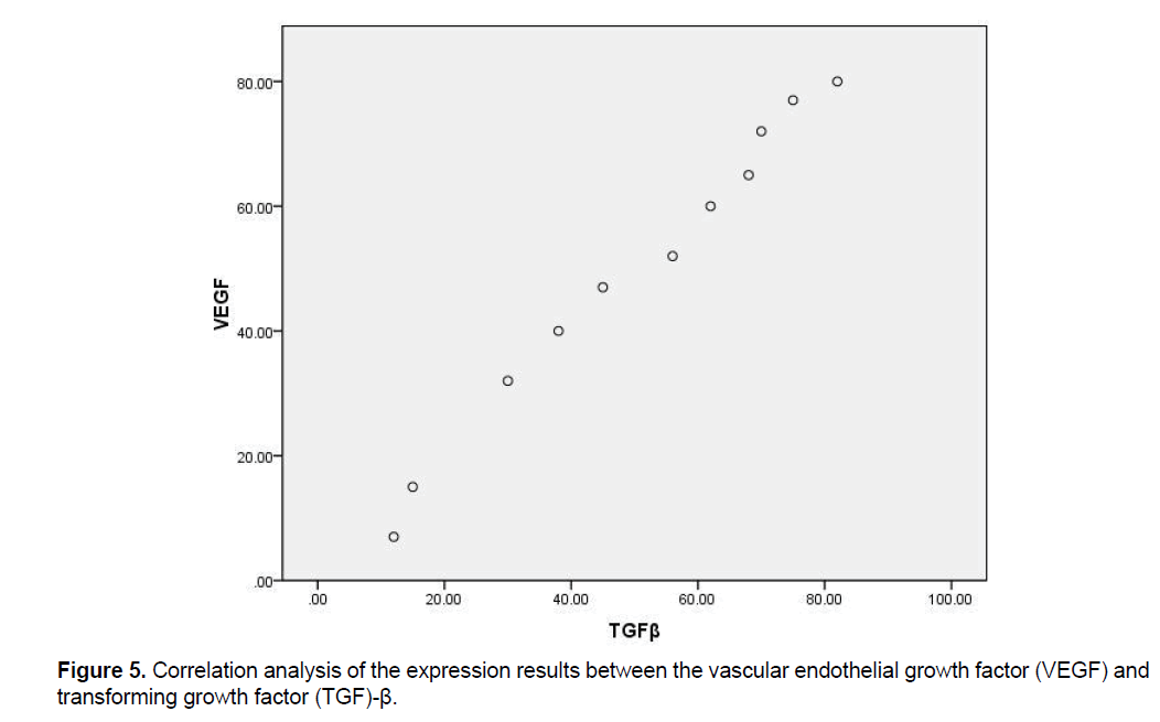
We analyzed the results of VEGF and TGF-β expressions by using Pearson correlation analysis, r=0.783, and expressions of the two factors were positively correlated (Figure 5).
4. Discussion
OA is a common chronic degenerative disease closely related to susceptible factors such as age, obesity, and excessive activity. As OA causes significant impairment to people’s lives, researchers have focused on this condition to help relieve patients’ pain [7]. OA is one of the most common forms of arthritis and includes intra-articular synovial and cartilage tissue lesions [8]. Patients with OA account for about 15% of the world’s population; the United States has about 200 000 patients with OA, and Europe has about 390 000 patients with OA. By 2020, these statistics for OA will double [9]. Pathological characteristics of OA include intra-articular synovial tissue fibrosis, articular cartilage osteophyte formation, sclerosis of the subchondral bone, labial hyperplasia around the joint edge, and intra-articular loose bodies [1]. Radiographs of OA show the corresponding changes: (1) A narrow joint space, (2) Sclerosis of the subchondral bone, (3) Osteophyte formation around peripheral joints, (4) cystic change in the subchondral bone, (5) Varus or valgus deformity, and (6) Subluxation of the joint. However, in a large number of clinical cases, a previous study found that the severity of the radiographic manifestation has no obvious correlation with clinical symptoms [10]; for example, some patients with severe OA on radiographs may have minimal clinical symptoms. Knee pain is often the main reason why patients seek treatment [11]. The pain occurs unilaterally or bilaterally when an individual with OA walks up and down the stairs during the early phase, especially when walking down the stairs, and joint swelling and varus or valgus deformity may occur. OA can be divided into primary OA and secondary OA. Primary OA has no obvious causes. Secondary OA is based on original disease development and there are many causes of the disease, including congenital dysplastic joints, pediatric joint disease, trauma, metabolic disease, and various types of articular cartilage inflammation. Articular cartilage injury is the main characteristic of OA, and the self-repairing capability of articular cartilage is limited. Articular cartilage injury accelerates disease progression, so early intervention for this injury is an effective method to alleviate joint disease progression [12,13].
| Comparison group | VEGF | TGF-β | ||
|---|---|---|---|---|
| χ2 | P value | χ2 | P value | |
| Group one and group two | 1.21 | >0.01 | 1.292 | >0.01 |
| Group one and group three | 10.402 | 0.0015* | 8.630 | 0.003* |
| Group one and group four | 16.871 | 0.000* | 14.545 | 0.000* |
| Group two and group three | 4.871 | >0.01 | 3.635 | >0.01 |
| Group two and group four | 10.001 | 0.001* | 8.121 | 0.004* |
| Group three and group four | 1.129 | >0.01 | 1.027 | >0.01 |
*P<0.01, differences between the two groups are statistically significant.
Table 5. Results of multiple comparisons of vascular endothelial growth factor (VEGF) and transforming growth factor (TGF)-β expressions among each group
Figure 3. Expression of transforming growth factor (TGF-ÃÆÃâÃâÃ
¸) in synovial tissue (SPX400). A: Negative expression of TGF-ÃÆÃâÃâÃ
¸
in synovial tissue; B: weak positive expression of TGF-ÃÆÃâÃâÃ
¸ in synovial tissue; C: positive expression of TGF-ÃÆÃâÃâÃ
¸ in synovial
tissue; and D: strong positive expression of TGF-ÃÆÃâÃâÃ
¸ in synovial tissue.
Note: Group one had no evidence of osteoarthritis; Group two patients showed mild osteoarthritis; Group three patients
had moderate osteoarthritis; Group four patients had severe osteoarthritis
To study the initiation and progression of knee OA, this study explored biological characteristics of knee OA through the expression of cytokines in articular synovial tissue. Numerous studies have found that angiogenesis of the joint synovial tissue, and cartilage and subchondral bone tissues play an important role in joint destruction [14]. However, the formation of blood vessels is not isolated, as it is regulated and induced by a series of cytokines. Among all the cytokines, VEGF and TGF-β play an important role in promoting angiogenesis and the development of OA by influencing each other. Based on this finding, we hope to determine the correlation between TGF-β, VEGF and OA development to improve treatment for OA and relieve patients’ pain.
4.1 Expression of VEGF in the synovial tissue of patients with OA
VEGF exists throughout tissues and organs. VEGF levels are relatively low in the normal population. Because the blood supply is rich in pathological tissue, VEGF levels are relatively high in tumor tissue, inflammatory lesions, and pathological tissues. Aside from lack of oxygen, hormonal and inflammatory factors can promote the expression of VEGF. The main physiological characteristics of VEGF are promotion of vascular endothelial cell proliferation, migration, capillary permeability, and blood capillary formation. Physical properties of the promotion of capillary formation lead to the occurrence and development of diseases that can be difficult to treat [15]. Lambert et al. reported that VEGF expression was more significant in an inflammatory area of synovial joint tissue than in a non-inflammatory area. To understand the effect of different synovial fluid on the original generation of articular cartilage cells, Landry et al. added normal knee articular cartilage cells with original generation to the joint fluid of patients with OA for culture; the VEGF level of synovial fluid was high [16]. VEGF can promote the formation of new blood vessels and change the permeability of blood vessels. Long-term effects include articular synovial tissue hypertrophy, and its pathological changes are closely related to the formation of new blood vessels [17]. Another study reported that the VEGF levels have a positive correlation with blood sedimentation rate, C-reactive protein level and swelling of the joints [18]. In the clinical setting, VEGF level of joint fluid indicates the activity of knee OA.
By comparing the experimental group and control group, we found that the joint synovial tissue of the experimental group had new blood vessels. Moreover, when the severity of the joint synovial tissue lesion became worse, we observed an inflammatory reaction and vascular proliferation, which plays an important role in the destruction of synovial tissue lesions. By evaluating the VEGF expression of articular synovial tissue using the immunohistochemical method, we found that the VEGF was mainly expressed in vascular endothelial cells and the cytoplasm of synovial membrane cells. VEGF expression among the study groups was different, and a comparison between the experimental and control groups showed a difference. Based on our findings and those of numerous reports, we think that normal physiological environment changes in the joint space are an important cause of OA [19]. New blood vessels in the synovial tissue of patients with arthritis and inflammatory cell proliferation provide a favorable environment; however, inflammatory cells can promote the secretion of VEGF and VEGF can increase the permeability of blood vessels, leading to intravascular macromolecular substances, such as collagen and fibrin seeping to the outside of the blood vessels, and an imbalance of osmotic pressure in the articular cavity [20]. Secondary vascular endothelial cell migration, vascular endothelial cell degradation, and endothelial cell proliferation by VEGF increase the severity of arthritis [21].
VEGF expression was significantly higher in osteoarthritic synovial tissue than in normal articular synovial tissues, and pathological changes of new blood vessel expression was significantly higher in osteoarthritic synovial tissue than in normal articular synovial tissue. These findings indicate that the high VEGF expression in knee joint synovial tissue is positively associated with the degree of pathological changes, and the VEGF level of joint fluid indicates the activity of knee OA.
4.2 TGF-β expression in the synovial tissue of patients with OA
TGF-β is an important growth factor, and it plays an important role in regulating cell proliferation and differentiation [22]. Through interaction with receptors, TGF-β exerts its biological effects, such as the repair of articular cartilage, reconstruction of bone, and wound repair. TGF-β can contribute to the proliferation and migration of endothelial cells, and promote the formation of new blood vessels.
In the immunohistochemical detection of TGF-β expression in arthritic synovial specimens, we found TGF-β in vascular endothelial cells, mesenchymal cells, and the cytoplasm of epithelial cells. In the mild, moderate, and severe groups, the rate of TGF-β expression was significantly different overall. In addition, the more capillary tissue expression in the synovial, the heavier the synovial tissue lesion is which increases the TGF-β expression. There were significant differences among multiple comparisons of the mild, moderate, and severe groups. This finding demonstrates that the TGF-β expression has a close relationship with pathological changes of osteoarthritic synovial tissue and as the degree of osteoarthritic synovial tissue worsens, the TGF-β expression will increase. According to reports worldwide, TGF-β has an indirect relationship with the production of capillaries of knee OA synovial tissue; moreover, it can induce the generation of new capillaries indirectly, rather than directly promoting synovial neovascularization formation [23]. However, the molecular mechanism needs to be researched further.
In our study, we found that TGF-β expression is significantly higher in knee arthritic synovial tissue than in normal articular synovial tissue. TGF-β expression is also higher in new blood vessels of synovial tissue. A high TGF-β expression is closely related with osteoarthritic synovial membrane changes [24]. However, previous reports have indicated that TGF-β has an indirect role in the formation of new blood vessels, so it should not be a reliable clinical diagnostic index of knee arthritic synovial tissue [25].
4.3 Relationship between TGF-β and VEGF expressions in knee OA synovial tissue
VEGF expression plays an important role in the formation of blood vessels, and it is the central regulating factor in synovial tissue angiogenesis. VEGF adjustment is not isolated, as it involves many factors in the formation of blood vessels [26]. TGF-β is a multi-function biological regulator, and in different cellular organizations, it plays a different role: promotion and inhibition. Many scholars believe that TGF-β has an important role in regulating VEGF expression. Studies have shown that the TGF-β is a chemokine for fibroblasts, monocytes, and neutrophils, and it promotes the direct release of vascular growth factors; therefore, VEGF expression increases simultaneously and TGF-β promotes vascular differentiation and maturation indirectly. Other reports have indicated that TGF-β has a dual function for VEGF, and the function has a close relationship with its level. Pepper et al reported that TGF-β has an obvious inhibitory effect on endothelial cells in terms of generation, development, and maturity at a higher concentration and dosage, but it can promote the growth and maturity of endothelial cells at a low dosage and concentration [27].
5. Limitations of Our Study
Through this experimental study, we found that the expression of TGF-β and VEGF had a significant correlation in knee osteoarthritic synovial tissue lesions. However, we need to consider that the humans are walking animals and that their living environment changes. There is still a difference in the organizational structure and internal environment in knee joints and these results still needs further validation in a larger series of patients.
This study experiment showed that the pathological changes of osteoarthritic synovial tissue and the expression of TGF-β and VEGF had an apparent correlation. TGF-β and VEGF offer new therapeutic targets, but their pathological mechanism in OA is not yet clear. Further research is needed to confirm the mechanism of action to help us develop more effective treatments.
6. Conclusion
We found that TGF-β and VEGF are simultaneously involved in the development of OA, and TGF-β and VEGF have similar high expression. TGF-β can activate VEGF, and VEGF can promote the formation of microvascular development; thus, TGF-β and VEGF have a role in OA development.
References
- Lambert C, Mathy-Hartert M, Dubuc JE, et al. (2012). Characterization of synobial angionenesis in osteoarthritis patients and its modulation by chondroitin sulfate. Arthritis Res Ther. 14: 1-11.
- Ludin A, Sela JJ, Schroeder A, et al. (2013). Injection of vascular endothelial growth factor into knee joints induces osteoarthritis in mice. Osteoarthritis Cartilage. 21: 491-497.
- Ropert S, Vignaux O, Mir O, et al. (2011). VEGF pathway inhibition byanticancer agent sunitinib and susceptibility to atherosclerosisplaque disruption. Invest New Drugs. 29: 1497-1499.
- Xu Y, Yuan L, Mak J, et al. (2010). Neuropilin2 mediates VEGFC induced lymphatic sprouting together with VEGFR3. J Cell Biol. 188: 115-130.
- Chen Y, Dawes PT, Mattey DL. (2012). Polymorphism in the vascularendothelial growth factor A (VEGFA) gene is associated withserum VEGFA level and disease activity in rheumatoid arthritis differential effect of cigarette smoking. Cytokine. 58: 390-397.
- Outerbridge RE. (1964). Further studies on the etiology of chondromalacia patellae. J Bone Joint Surg Br. 46: 179-190.
- Patel K, Raut V. (2011). Patella in total knee arthroplasty: To resurface or not to –a cohort study of staged bilateral total knee arthroplasty. Int Orthop. 35: 349-353.
- Gaëlle Clavel, Natacha Bessis, Delphine Lemeiter, et al. (2007). Angiogenesis markers (VEGF, soluble receptor of VEGF and angiopoietin-1) in very early arthritis and their association with inflammation and joint destruction. Clin Immunol. 124: 158-164.
- Lee YT, Shao HJ, Wang JH, et al. (2010). Hyaluronic acid modulates gene expression of connective tissue growth factor (CTGF), transforming growth factor-β (TGF-β) and vascular endothelial growth factor (VEGF) in human fibroblast like synovial cells from advanced stage osteoarthritis in vitro. J Orthop Res. 28: 492-496.
- Song JG, Han JH, Kwon JH, et al. (2015). Radiographic evaluation of complete and incomplete discoid lateral meniscus. Knee. 22: 163-168.
- Cakucg AL, Domiciano DS, Fuller R, et al. (2011). Osteoarthritis: Can anticytokine therapy play a role in treatment. Clin Rheumatol. 29: 451-455.
- Oshimori N, Fuchs E. (2012). The harmonies played by TGF-β in stem cell biology. Cell Stem Cell. 11: 751–764.
- Ropert S, Vignaux O, Mir O, et al. (2011). VEGF pathway inhibition by anticancer agent sunitinib and susceptibility to atherosclerosis plaque disruption. Invest New Drugs. 29: 1497-1499.
- Van den Berg WB. (2011). Osteoarthritis year 2010 in review: Pathomechanisms. Osteoarthritis Cartilage. 19: 388-341.
- Xu Y, Yuan L, Mak J, et al. (2010). Neuropilin2 mediates VEGFC induced lymphatic sprouting together with VEGFR3. J Cell Biol. 188: 115-130.
- Landry JP, Fei Y, Zhu X, et al. (2013). Discovering small molecule ligands of vascular endothelial growth factor that block VEGF KDR binding using label free microarray based assays. Assay Drug Dev Technol. 11: 326-332.
- Beamer B, Hettrich C, Lane J. (2010). Vascular endothelial growth factor: An essential component of angiogenesis fracture healing. HSS J. 6: 85-94.
- Tahara A, Tsukada J, Tomura Y, et al. (2011). Vasopressin induces human mesangial cell growth via induction of vascular endothelial growth factor secretion. Neuropeptides. 45: 105–111.
- Johnson K, Zhu S, Tremblay MS, et al. (2012). A stem cell-based approach to cartilage repair. Science. 336: 771-721.
- Poh CK, Shi Z, Lim TY, et al. (2010). The effect of VEGF functionalization of titanium on endothelial cells in vitro. Biomaterials. 31: 1578–1585.
- Neve A, Cantatore FP, Corrado A, et al. (2013). In vitro and in vivo angiogenic activity of osteoarthritic and osteoporotic osteoblasts is modulated by VEGF and vitamin D3 treatment. Regul Pept. 184: 81–84.
- Dong LQ, Yin H, Wang CX, et al. (2014). Effect of the timing of surgery on the fracture healing process and the expression levels of vascular endothelial growth factor and bone morphogenetic protein-2. Exp Ther Med. 8: 595-599.
- Oshimori N, Fuchs E. (2012). The harmonies played by TGF-β in stem cell biology. Cell Stem Cell. 11: 751–764.
- Young DA, Bui C, Barter MJ. (2012). Understanding CpG methylation in the context of osteoarthritis. Epigenomics. Epigenomics. 4: 593–595.
- Wu XY, Wu XP, Luo XH, et al. (2010). The relationship between the levels of gonadotropic hormones and OPG, leptin, TGFβ and TGF-β2 in Chinese adult women. Clin Chim Acta. 411: 1296-1305.
- Li X, Li N, Ban C J, et al. (2011). Idiopathic pulmonary fibrosis in relation to gene polymorphisms of transforming growth factor-β1 and plasminogen activator inhibitor. Chin Med J. 124: 687-690.
- Mori S, Fuku N, Chiba Y, et al. (2010). Cooperatibe effect of serum 25-hydroxyvitamin D concentration and a polymorphism of transforming growth factor-βgene on the prevalence of vertebral fractures in postmenopausal osteoporosis. J Bone Miner Metab. 28: 446-450.

Open Access Journals
- Aquaculture & Veterinary Science
- Chemistry & Chemical Sciences
- Clinical Sciences
- Engineering
- General Science
- Genetics & Molecular Biology
- Health Care & Nursing
- Immunology & Microbiology
- Materials Science
- Mathematics & Physics
- Medical Sciences
- Neurology & Psychiatry
- Oncology & Cancer Science
- Pharmaceutical Sciences


