Enhancement of Coronary Artery Blood Vessels Using Digital Subtraction Angiographic Technique
A Umarani
A Umarani*
Department of Electronics and Instrumentation Engineering, K.L.N. College of Engineering, Madurai- 625001, Tamilnadu, India.
- Corresponding Author:
- E-mail: duraiumarani@gmail.com
Received Date: April 01, 2016; Accepted Date: April 20, 2016; Published Date: April 27, 2016
Citation: Umarani A. Enhancement of Coronary Artery Blood Vessels Using Digital Subtraction Angiographic Technique. Electronic J Biol, 12:3
Abstract
Arteriosclerosis a widespread disease of the arteries. It is a kind of arteriosclerosis that causes coronary artery disease (CAD) in the form stenosis that is narrow downing of arteries. The X-ray angiography images suffer from background artefacts which obscure the visualization of the image to identify the stenosis. The artefacts must be eliminated to have an improved visualization of the blood vessels. The main aim of the proposed method is to eliminate the background artefacts present in the image subjected to investigation to identify the stenotic lesion in the pathway of the blood vessels by a technique named Temporal Mask Mode Digital Subtraction Angiography (TMDSA). The subtracted image shows the elimination of background structures, making the arteries highly visible. The performance is evaluated by estimating signal to noise ratio (SNR) and contrast to noise ratio (CNR). The feasibility of subtraction of coronary angiography images improves the interpretation of stenosis.
Keywords
Coronary angiography, Digital subtraction angiography, Coronary arteries, Coronary stenosis
1. Introduction
The leading cause of death worldwide is Coronary artery disease (CAD). The heart muscle become hardened and narrowed due to the build-up of plaque (fat deposits) on their inner walls due to which the blood supply to the coronary arteries gets restricted and it is termed as arteriosclerosis. Blood flow is reduced and the heart muscle does not receive sufficient oxygen due to the reduction of blood flow. When the plaque increases in size, the interior of the arteries, the lumen, gets narrower (stenosis) and less blood can flow through eventually. A blood clot develops at the site of the plaque and suddenly cuts off most or all the blood supply, which results in a myocardial infarction, causing permanent damage to the heart muscle [1]. The x-ray arteriography or X-ray angiography are used to study the lumen impairment. Since the contrast of the fluoroscopic image is limited by x-ray scatter in the patient, the blood vessels are not clearly visualized to identify the stenosis. Hence the In order to perform examination that are generally referred to as digital subtraction angiography (DSA) the injection of contrast material is combined with the acquisition of digital fluoroscopic images and realtime subtraction of pre-and post-contrast images to perform examinations.
2. Literature Review and Problem Formulation
To reduce the motion artefacts in coronary angiography, several methods have been proposed. First an overview of DSA technical principles was presented by Harrington et al. [2]. The DSA development and its uses were proposed by Jeans [3]. Analysis of image registration technique and its application in digital subtraction angiography was proposed by Taleb and Jetto [4]. The process of spatially aligning of the identical two or more images taken at varying times and from unique viewpoints is known as image registration. The elimination of artefacts in DSA images based on a simple rigid model using a successive approximation algorithm is presented by Funakami et al. [5]. In this approach, combined invariant-based similarity method based on edge detection was used to eliminate the presence of background artefacts in DSA. Due to the vessel overlap, multiple projections are necessary to evaluate the coronary artery. To overcome this problem Chen and Carroll [6] proposed a technique of constructing of 3-D coronary artery from 2-D DSA image sequences employed with some bifurfication which reduces the overlapping area and automatically extracts coronary artery but this required number of bifurfication and does not consider the noise effects. A dynamic feature extraction of coronary artery motion in DSA image sequences was proposed by Puentes et al. [7]. In this issue the local and global features of coronary arteries are extracted to identify the motion patterns. The displacements of the artery were easily described using this feature extraction method. Meijering et al. [8] proposed a fully automatic method to reduce the motion artefacts in digital subtraction angiography. Further Taleb et al. [1] extended his research in developing an image registration technique with evaluation based on a 3-D spacetime motion in DSA. In this approach, for selecting the moving points belonging to a moving structure a 3-D space-time motion detection algorithm was used. The degradation of the image is eliminated by the above approach. This approach does not concentrate on gray level variations or distortion even though it does not affect any diagnostic value; therefore, occasionally it leads to misinterpretation of data. Image registration of DSA using an invariant approach was presented by Bentoutou et al. [9].The efficient 3D registration and refinement technique for DSA was focused by Kwon et al. [10]. Bentoutou and Taleb [11] extended his research in DSA by developing a 3-D space-time motion detection based on invariant image registration approach.
The artefacts in DSA are caused mainly due to the movement of the patient, and therefore Shechter et al. [12] focused on motion correction method in X-ray angiography images. This work is further extended by developing a comparative study on motion correction strategy done by Kumar et al. [13]. The extraction of vessels from DSA images based on the scan line tracking method is proposed by Zou et al. [14]. Further, this study report on shifty ahead detection schemes for continuation points in the vascular tree, but the drawback is it leads to discontinuities of vessels Yamamoto et al. [15] concentrated on the complete description on DSA for coronary arteries. Ionaseca et al. [16]; Bousse et al. [17] focused on motion compensation caused during image acquisition in digital subtraction angiography. Temiz et al. [18] presents DSA for persistent trigeminal artery for a cerebral angiogram. Umarani and Asha [19] presents automatic digitization and subtraction of images to enhance the blood vessels. Tanaka et al. [20] proposed a subtraction method considering the coronary calcium deposit along the arteries. This review describes a temporal mask time mode digital subtraction angiography (TMDSA) technique to reduce the motion artefacts. The technique was applied to real time data sets for an enhanced visualization of blood vessels and for the discussion of coronary stenosis.
There is no research done to eliminate the artefacts in the coronary angiography images considering the effect of noises. During clinical subtraction, the pixel value gets subtracted, but the noise adds along with it creating an obscuration in viewing the vessels. Hence the process of subtracting two images has the unfortunate consequence of producing a noisier subtracted image, and it is taken into consideration during the diagnosis. Considering all the above issues the proposed method, removes the artefacts from the coronary angiography images with effect to noise distributions. Therefore, the purpose of the proposed method is to eliminate the artefacts by employing a technique named Temporal Mask time mode digital subtraction Angiography (TMDSA) to visualize the blood vessels clearly. For a real time, it is really hard to use the DSA technique to the coronary arteries because of the severe motion artefacts caused by cardiac motion and respiration. Consequently an image-processing technique can be employed to overcome this problem. Since the clinical subtracted images possess noise during image subtraction images, the proposed method of image subtraction overcomes this drawback by eliminating the noise before the images are subjected to digital subtraction. Therefore, only the denoised images are considered as input for the subtraction process. Therefore the Time Mode Digital Subtraction Angiography (TMDSA) technique is used to reduce the motion artefacts in X-ray Angiography images.
3. Materials and Methods
The images are acquired on a Toshiba C arm machine which is equipped with an image intensifier and a digital flat-panel detector. The scans are performed at the rate of 15 to 30 frames per second (fps).The data sets were saved in the Digital Imaging and communication in medicine (DICOM) format, a medical standard in most of the imaging modalities for transfer of images, movies and other diagnostic data. The quality of the DICOM image must be improved for early diagnosis of disease. The data sets are collected from the Government Rajaji Hospital, cardiology section, Madurai, India. For optimal visualization, multiple projections are obtained during angiography of native vessels and grafts, including Left anterior oblique (LAO), Right anterior oblique (RAO), frontal, lateral and cranial and caudal views. Twenty data sets are acquired with a C -Arm Machine. A tube voltage of 85 kV and a tube current of 450 to 500mA are used for both scanners. As a result of image acquisition, a series of 512 × 512 16 bit gray scale images with 256 different gray levels (0-255) are produced.
4. Outline of the Proposed Method
Visualization of blood vessels that has to be diluted with a radiographic contrast media requires subtracting the high-contrast structures from the image. The subtraction image can be enhanced using display windowing techniques routinely employed for angiography images only when the high contrast structures have been cancelled. These contrast enhancement techniques are ineffective without some form of subtraction, because the high contrast objects, e.g., bone/soft-tissue and air/softtissue interfaces will obscure the lower contrast blood vessels containing to dilute iodine. Temporal subtraction, energy subtraction and hybrid subtraction prevail for soft-tissue and bone cancellations are three methods of subtraction. Temporal subtraction, familiar to radiologists, requires an image before administration of contrast (the mask) which is then subtracted from all subsequent post-contrast images (Phase).Complete cancellation of soft-tissue and bone is achieved with this method, leaving only the iodinated blood vessels by assuming there has been no subject motion between the two images. Thus, the proposed method comes under temporal subtraction method, and the subtraction process is carried out considering the mask image and the phase image by a technique named Temporal Mask time mode digital subtraction Angiography (TMDSA).
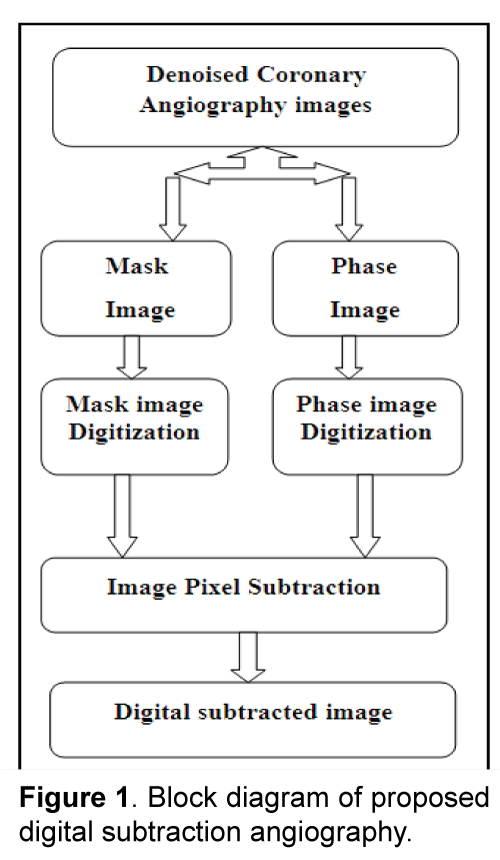
The proposed block diagram is shown in Figure 1. The process flow is as follows, according to the capturing rate the angiography video images are converted into image slices. For image subtraction analysis two images specifically mask and phase images are considered with no deviation in frames, and in the pixel values and the images are acquired at a single time. Mask image is the image obtained before the injection of contrast agent and phase image is obtained after injecting the contrast media. An image consists array of elements and dimensions. Each element represents as pixels. An image consist array of elements and dimensions. Each element represents as pixels.
The physical point in an image or the smallest addressable element in a display device is the pixel in digital imaging. So it is the tiniest controllable element of a picture represented on the screen. A pixel physical coordinates corresponds to its address. The intensity of each pixel is variable. The gray scale images are represented in terms of gray shades, and the intensity value varies from 0- 255 and entirely it has 256 gray values. For the subtraction process, both the images are subjected to digitization to obtain the pixel values. Subsequently the pixel value of the mask image is subtracted from the pixel value of the phase image. The process undergoes linear and logarithmic subtraction to obtain a digital subtracted angiography image.
As a result, the purpose of the proposed method is to eliminate the artefacts by employing a technique named Temporal Mask time mode digital subtraction Angiography (TMDSA) to visualize the blood vessels clearly. In real time, due to the severe motion artefacts caused by cardiac motion and respiration, it is very difficult to apply the DSA technique to the coronary arteries. Thereby an image processing technique can be employed to overcome this problem. Since the clinical subtracted images possess noise during image subtraction images, the proposed method of image subtraction overcomes this drawback by eliminating the noise before the images are subjected to digital subtraction. Therefore, only the denoised images are considered as input for the subtraction process. The technique can be applied to real time data sets with reducing motion artefacts caused by various factors for an enhanced visualization of blood vessels and for the treatment of coronary stenosis. The quality of the subtracted image is estimated by computing, signal to noise ratio (SNR) and contrast to noise ratio (CNR). The image processing analysis is done in the National instrument (Ni) Lab VIEW environment.
5. Results and Discussion
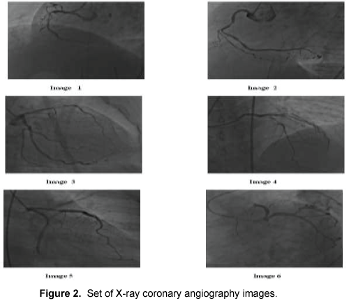
The performance of the proposed method was tested on a real data set collected from the Government Rajaji Hospital, cardiology section, Madurai, India. Toshiba C-Arm machine are used as X-ray imaging and the total frame varies according to the frame rate 30 frames per second, 15 frames per second, 7.5 frames per sec, 375 frames per second, 1.88 frames per second. Iodine is used as the contrast agent to acquire the vessel-enhanced image (Phase) and the actual dose of the iodine contrast varies on the patient and anatomy of the individual being imaged. The data set used for the evaluation consist of sequences of 512x512 images and has a gray value resolution of 8bit and the grey values varied from 0-255, i.e. 256 gray level. Figure 2 illustrates a set of coronary artery angiography images. The image quality is most evenly degraded by image noise, and it must be eliminated in both the phase image and mask image before it is subjected to subtraction.
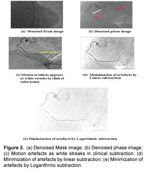
The proposed method aims to eliminate the background artefacts that are the bones and tissues to visualize the coronary artery blood vessels clearly to help the clinician in the proper diagnosis of coronary artery disease. The proposed technique uses a temporal mask time mode digital subtraction Angiography (TMDSA) techniques to eliminate the artefacts. To implement the proposed technique, a de-noised mask image and a de-noised phase image are considered as shown in the Figures 3a and 3b. The mask and the phase image selected should be from the identical subject, and the image should be acquired in the identical period. If the images acquired at various time for the equivalent subject is considered for image subtraction, then it leads to pixel shifting, and the subtraction process will become very difficult. Considering the images of the same subject and using the de-noised coronary angiography the proposed technique is performed. Figure 3c shows the clinical subtraction carried out in the clinical environment. Here the white streaks appear as one of the artefacts. Therefore, the artefacts are removed by linear and logarithmic subtraction. Figures 3d and 3e shows the minimization of artefacts by linear subtraction and Logarithmic subtraction. Both the images are digitized using image digitization techniques. Then the subtraction is performed between the digitized pixel values of mask image and phase image. The digitized value of the mask image is subtracted from the digitized value of the phase image and therefore pixel-by-pixel subtraction is made.
During subtraction, the pixel value of both the image gets subtracted, leaving a subtracted output pixel value. The technique undergoes two subtractions namely linear and logarithmic subtraction in linear subtraction mathematical subtraction is computed whereas in logarithmic subtraction the logarithmic values for each pixel value is calculated and then the subtraction is performed. In both the subtraction, the subtracted pixel values may appear as positive value or negative value as illustrated in Figure 4. If the subtracted output pixel values are lesser than zero i.e., negative values are attenuated to zero and if the subtracted output pixel value is greater than 255 then that values are attenuated to 255. The common pixel value of the images gets subtracted indicating a zero pixel value which means that the background structures i.e., bones and tissues get eliminated leaving behind the enhanced coronary blood vessels.
The patient should be immobile between the acquisition of the mask and phase images, otherwise the images will not be registered properly and motion artefacts, in the form of whitish streaks (shown in arrow mark), will be visible in the subtracted image during clinical analysis. This is caused due to the patient move and the artefacts must be eliminated for proper diagnosis of blood vessels. The clinical solution is to shift the mask image by a few pixels; to prevent the motion, before subtraction but these pixels shifting tend to be a trial-and-error process, involving a combination of shifts in different directions and by differing amounts and it leads to misinterpreting of data’s. Therefore, the proposed method of elimination of artefacts using pixel subtraction with linear and logarithmic subtraction is illustrated in Figures 3d and 3e. The qualitative analysis of the proposed DSA images shows a clear visualization of blood vessels than the clinical subtracted image. It is feasible to select a best subtracted image from a set of phase image of the same subject. For this purpose, automatic digitization and subtraction are done by using a mask image and a set of phase image. In this method after digitization, mask image is subtracted from all the frames of the phase image automatically Umarani and Asha [19]. Through this operation, the best quality of the image can be selected by estimating the SNR to visualize the blood vessels clearly.
The image quality is evaluated in terms of signal-tonoise ratio (SNR) and contrast to noise ratio (CNR). SNR is the ratio of the mean of intensity difference between the signal (foreground) and the noise (background) to the standard deviation of the noise, and it is often expressed in decibels (dB). The noise power (PN) can be taken as the variance of pixel values, but needs to be measured in a region within the image which is expected to have constant gray values and is large enough so that all significant variations are included in the noise measurement. The signal, or mean intensity (PS) of the image is characterized by the square of the mean pixel value of the entire image. To understand its significance, the noise (PS) should be compared to the average power or intensity of the signal (PS). The signal power is determined by the square of the average pixel value in the entire image and the noise power is determined from the variance within a region of interest (ROI) containing no features. Thus SNR is given by
SNR = 10 log10 (PS/PN) (1)
In medical imaging, the goal is to distinguish a foreground structure from the background. In such cases, it is better to estimate the contrast - to- noise ratio (CNR), rather than the SNR and the CNR is given by
 (2)
(2)
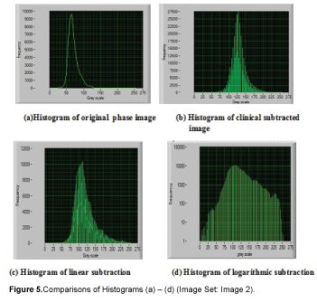
Where the signal, or average pixel value of the whole image, is replaced by the contrast, or the difference in the average pixel values of the foreground and background. The CNR ratio is equal to the difference in SNR for the foreground and background, respectively, since the noise is similar whether measured using the foreground or background pixels. The signals are the visualized blood vessels and the noises are the background regions in the medical images as shown in the Figure 3b. The signal the blood vessels and the noise the background are calculated in each image and the mean, standard deviation based on pixel intensity is computed to determine the SNR and CNR. The comparative DSA results are shown in Figure 5. From the qualitative analysis, it is found the blood vessels are clearly visualized in the proposed method than the clinical subtracted DSA image. The analysis can be interpreted using the image intensity graph called a histogram. A graph showing the number of pixels in an image at each distinctive intensity value found in the image is called the histogram. For an 8-bit grayscale image, there are 256 distinct possible intensities, and so the histogram will graphically display 256 numbers showing the distribution of pixels among those grayscale values.
The intensity distribution (histogram) of the image is shown in Figure 5. From the intensity curve, it is found, the denoised phase image illustrated in Figure 5a and clinical DSA image in Figure 5b and the linear subtraction as in Figure 5c, the gray values are not distributed over the entire scale and it is narrowed down and because of that the image is not that much clear to visualize the blood vessels. However, in the proposed method of logarithmic subtraction, the intensity graph illustrated in Figure 5d shows that the gray values are distributed to the entire gray scale value, and consequently the image quality gets improved than the clinical DSA images. The quantitative analysis of estimation of SNR and CNR to determine the quality of the image is given in the Table 1. It is found that the SNR of the logarithmic DSA images is greater than the SNR of the other DSA images and CNR is less than that of SNR, from which we tend to conclude that the proposed method of image subtraction had a more coherent image quality than the clinical DSA.
6. Conclusion
Thus, this new method is proposed to improve image quality using DSA. To visualize the coronary artery blood vessels clearly, first, the images are subjected to noise elimination and to the denoised images, the motion artefacts are expelled using a technique named Temporal Mask time mode digital subtraction Angiography (TMDSA). The put forward method has been evaluated on a clinical database of coronary artery Angiography images. The SNR and CNR for a set of coronary angiography image were determined and a comparison of qualitative and quantitative analysis is done for the clinical data set and the proposed data sets. From the experimentation, it has been observed that the proposed system is capable of eliminating the artefacts well on coronary angiograms. The results in terms of SNR and CNR are found to be comparable and superior to clinical approaches and deserve to be a reliable method for eliminating the artefacts in coronary angiograms.
References
- Taleb N, Bentoutou Y, Deforges O, et al. (2001). ‘A 3-D space-time motion evaluation for image registration in digital subtraction angiography’. Computer Medical Imaging Graphics. 25: 223-233.
- Harrington DP, Boxt LM, Murray PD. (1982). ‘Digital subtraction angiography: overview of technical principles’. American Journal of Roentgenology. 139: 781-786.
- Jeans WD. (1990). ‘The development and use of digital subtraction angiography’. British Journal of Radiology. 63:161-168.
- Taleb N, Jetto L. (1998). ‘Image registration for applications in digital subtraction angiography’. Control Engineering Practice. 6: 227-238.
- Funakami R, Hiroshima K, Nishino J. (2000). ‘Successive approximation algorithm for cancellation of artefacts in DSA images’. The Japanese Society of Medical Imaging Technology. 18:199-206.
- Chen SJ, Carroll JD. (2000). ‘3-D Reconstruction of Coronary arterial tree to optimize angiographic visualization’. IEEE Transaction on Medical imaging. 318-336.
- Puentes J, Roux C, Garreau M, et al. (2000). ‘Dynamic feature Extraction of coronary artery motion using DSA image sequence’. IEEE Transaction on Medical imaging. 857-871.
- Meijering EHW, Niessen WJ, Bakker J. (2001). ‘Reduction of patient motion artefacts in digital subtraction angiography: evaluation of a fast and fully automatic technique’. Radiology. 219: 288-293.
- Bentoutou Y, Taleb N, Mezouar MCE, et al. (2002), ‘An invariant approach for image registration in digital subtraction angiography’. Pattern Recognition. 35: 2853-2865.
- Kwon SM, Kim YS, Kim TS, et al. (2004). ‘Digital subtraction CT angiography based on efficient 3D registration and refinement’. Elsevier Computerized Medical Imaging and Graphics. 28: 391- 400.
- Bentoutou Y, Taleb N. (2005). ‘A 3-D space-time motion detection for an invariant image registration approach in digital subtraction angiography’. Computer Visual Image Understanding. 97: 30-50.
- Shechter G, Shechter B, Jon Resar R. (2005), ‘Prospective Motion Correction of X-Ray Images for Coronary Interventions’. IEEE Transactions on Medical Imaging. 24: 441-450.
- Kumar D, Shen D, Wei L, et al. (2007), ‘Motion correction strategies for interventional angiography images: a comparative approach’. IEEE. 497-500.
- Zou P, Chan P, Rockett P. (2009). ‘A Model-Based Consecutive Scanline Tracking Method for Extracting Vascular Networks From 2-D Digital Subtraction Angiograms’. IEEE Transactions on Medical Imaging. 28: 241-249.
- Yamamoto M, Okura Y, Ishihara M, et al. (2009). ‘Development of Digital Subtraction Angiography for Coronary Artery’. Journal of Digital Imaging. 22: 319-325.
- Ionaseca RI, Heiglb B, Hornegger J. (2009). ‘Acquisition-related motion compensation for digital subtraction angiography’. Elsevier Computerized Medical Imaging and Graphics. 33: 256-266.
- Bousse A, Zhou J, Yang G, et al. (2009). ‘Motion Compensated Tomography Reconstruction of Coronary Arteries in Rotational Angiography’. IEEE Transactions On Biomedical Engineering. 56: 1254-1257.
- Temiz O, Genchellac H, Unlu E, et al. (2010). ‘Digital subtraction angiography of a persistent trigeminal artery variant’. Diagnostic Interventional Radiology. 16: 245-247.
- Umarani A, Asha A. (2012). ‘A Novel Approach to Automatic Digitization and Subtraction of X-ray Image for Percutaneous Coronary Artery using Virtual Instrumentation’. European Journal of Scientific Research. 582-591.
- Tanaka R, Yoshioka K, Muranaka K, et al. (2013). ‘Iwate/ JP Subtraction coronary CT angiography with iterative reconstruction a feasibility study of coronary calcium subtraction’. European society of radiology. 1-23.

Open Access Journals
- Aquaculture & Veterinary Science
- Chemistry & Chemical Sciences
- Clinical Sciences
- Engineering
- General Science
- Genetics & Molecular Biology
- Health Care & Nursing
- Immunology & Microbiology
- Materials Science
- Mathematics & Physics
- Medical Sciences
- Neurology & Psychiatry
- Oncology & Cancer Science
- Pharmaceutical Sciences


