The Prevalence of Iron-Deficiency Anemia in Non-Pregnant Women of Reproductive Age [14-45] with Anemia in Marvdasht's Shahid Motahari Hospital in 2012-2013
Saranaz Jangjoo, Leila Hosseini
Saranaz Jangjoo1, Leila Hosseini2,*
1MD, School of Medicine, International Branch, Shiraz University of Medical Sciences,Shiraz, Iran;
2Assistant Professor, Faculty of internal medicine,School of Medicine, International Branch, Shiraz University of Medical Sciences,Shiraz, Iran.
Received Date: May 18, 2016; Accepted Date: June 16, 2016; Published Date: June 23, 2016
Citation: Jangjoo S, Hosseini L. The prevalence of iron-deficiency anemia in non-pregnant women of reproductive age [14-45] with anemia in Marvdasht's Shahid Motahari hospital in 2012-2013. Electronic J Biol, 12:3
Abstract
The present study was designed and conducted with the aim of determining the prevalence of irondeficiency anemia among non-pregnant women aged 15 to 45 years old and anemia visiting Marvdasht’s Shahid Motahari hospital in 2012-2013. The present study is a cross-sectional study that has been conducted with the aim of exploring iron-deficiency anemia in 200 non-pregnant women aged 14 to 45 with anemia. Serum iron test, Total Iron-Binding Capacity [TIBC] and ferritin blood test were done and the prevalence of iron-deficiency was measured. The data were analyzed using descriptive statistical tests and t-test. The results indicated that iron-deficiency anemia in individuals with anemia was 6.5%. The mean of the levels of hemoglobin, hematocrit, the number of red blood cells, Mean Corpuscular Volume [MCV], Mean Corpuscular Hemoglobin [MCH] and Mean Corpuscular Hemoglobin Concentration [MCHC] in individuals with iron-deficiency anemia was equal to 9.7 gr/dl, 34%, 4.7 mil/mm3, 73 fl, 21.9 pgr and 28.6%. Also the mean serum iron level was estimated as equal to 18.3 μg/dl, Total Iron-Binding Capacity [TIBC] as equal to 427.3 μg/dl and ferritin mean was estimated as equal to 6.8 μg/l in individuals with iron-deficiency anemia. It is necessary to use timely and appropriate treatment for all women of reproductive age that have clinical or laboratory symptoms of iron-deficiency anemia.
Keywords
Anemia; Iron deficiency; Women.
Introduction
One of the groups that are at risk of nutritional deficiency is women of reproductive age [14-45 years old]. The severity of the deficiency is different depending on the region. Women have special nutritional needs due to the periods of pregnancy, lactation and menstruation that experience in their life. Women’s socioeconomic level and their knowledge of nutritional needs are among important factors that can prevent malnutrition in women. Marriage and pregnancy at an early age, short time periods between multiple pregnancies and long lactation periods, malabsorption, bacterial, viral and parastic infections, poverty and high costs of food, inappropriate dietary habits and patterns, inappropriate nutrition, lack of use of a specific food and psychological condition especially in adolescent girls are other factors that make women susceptible to nutritional deficiency. The aforementioned factors are more prevalent in developing countries, compared with developed countries. Generally when the body faces with a deficiency, body’s reserves of a material are depleted and the concentration of that material is reduced in blood and tissues and the urinary excretion and its metabolites are reduced. In the next stage, dysfunctions appear in natural body functions and anatomic lesions specific to that disease appear. Nutritional deficiency can include the deficiency of calories, proteins, vitamins, iron and the like. What is discussed in this study is iron deficiency that is perhaps one of the main reasons for nutritional deficiency in most societies. Iron is one of the main elements in functioning of body cells and the iron required is different in different body tissues. The main role of iron in the body is carrying oxygen in the structural framework of hemoglobin. Anemia is in the form of reduction of hemoglobin concentration or the reduction of red blood cell concentration in the circulatory system that results in the reduction of oxygen transfer capacity [1-4]. Iron-deficiency anemia refers to the anemia in which the level of iron in the body is lower than the normal level or in other words, the level of iron in bone marrow is lower than the level that is required for production of red blood cells [1]. Iron deficiency is one of the most common types of nutritional deficiency in a way that 50 percent of the anemia in the world are attributed to iron deficiency and this deficiency results in the death of 841000 individuals in the world each year [1]. Other factors impacting the prevalence of iron deficiency are socioeconomic condition, sex and race in terms of death, Africa and some parts of Asia have 71 percent of the deaths in the world and North America has 1.4 percent of the diseases and deaths related to the iron deficiency [1-6]. In severe iron-deficiency anemia that is not accompanied by other diseases red blood cells are hypochromic and microcytic, plasma’s iron concentration is reduced, TIBC level is increased, serum ferritin concentration is low, serum protoporphyrin concentration is increased and the level of iron stored in the bone marrow is reduced. Unfortunately a combination of these laboratory findings is only visible when the iron-deficiency anemia is very severe or is not accompanied with other diseases such as malignant neoplasms and infections. Exploring these laboratory findings is also difficult when the patient has received iron or blood for treatment of anemia [3]. This study aims to determine the prevalence of iron-deficiency anemia among non-pregnant women [of reproductive age] with anemia to aid in creating appropriate policies for prevention and treatment. Timely diagnosis of this disease results in taking action for necessary treatment and prevention for improving mothers’ health. In this regard, it is necessary to identify the prevalence level of this disease in the society [7]. This study aims at determining iron-deficiency anemia among non-pregnant women of productive age that have anemia to aid in creating appropriate policies for prevention and treatment.
Methodology
Study method
This study is conducted in Marvdasht which is one of the counties in northern part of Fars province of Iran with a population of 310540 individuals [based on the estimation of Iran’s Center for Statistics in 2011]. The population of the study is all non-pregnant women aged 14 to 45 with anemia that visited internists in Marvdasht’s Shahid Motahari hospital in 2012-2013 and were sent to laboratory for complementary tests, without considering marital status, place of residence [the district or neighboring villages], occupation, socioeconomic and cultural status, number of pregnancies and number of children. The present study is a cross-sectional study that is conducted with the aim of exploring iron-deficiency anemia in women of reproductive age with anemia in the county Marvdasht during 2012 and 2013. The same size was 200 individuals that were selected using convenience sampling based on the time visiting the hospital and considering the conditions of the study samples. It should be pointed out that participation in the study was completely voluntary and consent form was completed by each individual. In the case of an individual’s the lack of wiliness to participate, the next individual was asked to cooperate. A good cooperation of those visiting the hospital was seen. Identification code was used instead of registering individuals’ particulars on the blood samples and the questionnaires were numbered using this code too in order to observe the confidentiality of the information and moral aspects of the study.
Sample size determination
If the prevalence is 50 percent and the maximum difference between the estimation of the disease and the real value is 5 percent and the accuracy is 7 percent, the sample size for estimation of the prevalence is 200 individuals.
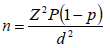
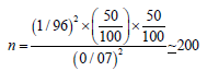
Where n is the sample size, p is the maximum prevalence level and d is accuracy.
The completion of the questionnaire
The data collection tool was questionnaire (Appendix) and some information was obtained thereby. The questionnaire was consisted of four sections: the first section was the medical-health history that included variables such as age, marital status, height, weight, occupation, age in which menstruation began, number of pregnancies, number of children, place of residence, history of blood disease, familial history of a special disease, the reason and problem for which the patient has visited the clinic of Marvdasht’s Shahid Motahari hospital and history of other diseases. The second section included clinical symptoms such as feeling of weakness, tiredness, dizziness, dyspnea, palpitations, pale skin, difficulty in concentration or the lack of clinical symptoms. The third section included medications and their consumption doses or the treatments the patients received in previous months in the case of being diagnosis with anemia. The fourth section included laboratory findings such as CBC, serum iron, ferritin and Total Iron-Binding Capacity [TIBC]. It should be pointed out that the validity and scientific authenticity of this questionnaire is based on the books and papers in Iran and was finally verified by the internists. The information related to the questionnaires was collected by two employees [having bachelor’s degree in laboratory sciences] of the laboratory of Marvdasht’s Shahid Motahari hospital and with face to face interview with the members of the sample.
The criteria for selecting the individuals in the sample included:
1. Age of 14 to 45 years
2. Lack of history of chronic diseases [cardiovascular, renal, metabolic and etc.] and inflammatory diseases based on the view of the medical center’s physician.
3. Not being pregnant
4. Women with anemia that is defined as hemoglobin lower than 12 g/dl [1].
5. Women who are not in their period during this study.
6. The lack of recent consumption of iron medicine.
Sample collection and test
After the completion of the questionnaire, as the iron level in the body varies during the day and the best time for sample collection is the morning in which the level of iron is natural and does not increase or decrease, all the samples were collected in 8 to 10 AM. 2 ml of venous blood were taken by venoject and in tubes containing Ethylen Diamine Tetra-Acetic Acid [EDTA] and hematological parameters were measured using Sysmex cell counter device [kx21, Japan] model kx21 in the laboratory of Marvdasht’s Shahid Motahari hospital. These parameters included hemoglobin, hematocrit, red blood cell count, white blood cell count, mean corpuscular volume [MCV], Mean Corpuscular Hemoglobin [MCH] and Mean Corpuscular Hemoglobin Concentration [MCHC]. Also, 5 ml of venous blood was collected in ironfree and acid-washed tubes for complementary tests i.e. serum iron, total iron binding capacity and ferritin [for preparing iron-free tubes, the tubes are put into choloridric acid 50 percent and then are put into double-distilled water and are washed with distilled water several times and are finally paced in room temperature to dry. The blood serums were separated immediately and serum iron and total iron binding capacity tests were done at the same day in less than 24 h in the biochemistry section of in the laboratory of Marvdasht’s Shahid Motahari hospital by Furno autoanalyzer model CA180[CA180, Japan] and using Parsazmun kit. And serum ferritin was measured using ELISA device, model Antus 2020 [Antus 2020, Netherlands] and using Padtan Elm kit in the biochemistry section of in the laboratory of Marvdasht’s Shahid Motahari hospital.
Measuring iron
Measuring iron is done using photometric method with wavelength of 600 nm by Furno autoanalyzer. Serum iron is freed from transferrin using pH reduction. And proteins and apotransferr are eliminated using precipitation and with centrifugation. Trivalent iron is reduced to divalent iron with ascorbic acid. Divalent iron forms a color complex with a chromogen. This group bonds with active iron and forms a pentagon ring structure that produces blue color. Ironchromogen comlex has a high absorption and is in proportion to the light absorption of iron.
Transferin(Fe2+) + HCL - trichoroacetic Acid → Transferin(Fe3+)2
Transferin(Fe3+)2 2Fe2+ + Transferin

Total iron binding capacity measurement
First, ammonium ferric citrate in appropriate quantity is added to the serum so that the serum iron places on transferrin are completely saturated. Then we wait for a while so that the trivalent iron bonds with transferrin to the maximum level. The reclaimed trivalent iron that is not bonded with transferrin is absorbed by adding magnesium carbonate powder and buffer. The mixture is centrifuged and the separated iron is measured. One thing that is important to be considered in measuring iron is that all tubes and glass instruments should be put into choloridric acid and washed with distilled water so that the tubes become free of iron.
Ferritin measurement
The kit used for quantitative measurement of ferritin is designed based on measuring immunoenzymatic reaction on solid phase. In this system, two antibodies are used that identify distinct antigenic indices on ferritin molecule. First, the standards, serum controls and the patients’ samples are added to the polystyrene holes covered with ferritin polyclonal antibodies. During the first incubation, the ferritin in the samples is attached to the anti-ferritin antibodies and other materials exit the system. After washing after the second incubation and washing with adding substrate 3,3',5,5'-Tetramethylbenzidine, a blue enzyme is created that is directly related to the ferritin concentration. The severity of the color is measured with spectrophotometer at 450 nm wavelength. For determining the ferritin concentration, first the standards’ light absorption curve is plotted based on ferritin concentration and then the samples’ ferritin concentration is determined from the standard curve.
Data analysis method
The coded information was analyzed using appropriate statistical methods after data collection and blood tests. The data were analyzed with the aid of computer and using SPSS software version 20 [the data will be considered significant with P<0.05]. Descriptive statistics and t-test were used for data analysis.
Results
In 200 women with anemia that were explored in this study, 4.5 percent were 14 to 17 years old, 6 percent were 18 to 21 years old, 14 percent were 22 to 25 years old, 15.5 percent were 26 to 29 years old, 18 percent were 30 to 33 years old, 8.5 percent were 34 to 37 years old,13 percent were 38 to 41 years old and 20.5 percent were 42 to 45 years old. Thus, the women aged 42 to 45 had the highest percentage and individuals aged 14 to 17 had the lowest percentage of the sample (Table 1). The anthropometric particulars of the studied sample are given in Table 2. The height mean in the whole sample was 163.55 ± 5.98 cm, the minimum height was 146 cm and the maximum height was 180 cm. The mean weight of the whole sample was 65.71 ± 10.19 kg, the minimum weight was 40 kg and the maximum weight was 100 kg. 76 percent of the individuals in the sample were married and 22.5 percent were single. The mean of the age in which menstruation began in them was 13.46 ± 1.31 years. 2.76 ± 1.27 was the mean of the number of pregnancies in married individuals [155 individuals=]. Regarding the number of abortion, in 155 individuals that had history of pregnancy 23.22 percent had the history of abortion. And one individual has the history of giving birth to a twin. The mean of the number of children was 2.47 ± 1.11. In terms of occupation, most of the individuals in the sample [86 percent] were just doing their housework [housewives] and 6.7 percent were employed. A series of clinical symptoms appear after anemia and some of them like feeling of weakness, tiredness, dizziness, dyspnea, palpitations, pale skin, difficulty in concentration and tingling of feet and hands are common symptoms can help in diagnosing iron-deficiency anemia. 36.5 percent of the women [73 individuals] had feeling of weakness, 41 percent [82 individuals] had the feeling of tiredness, 22.5 percent [45 individuals] had the feeling of dizziness, 18 percent [36 individuals] had dyspnea, 19 percent [38 individuals] had palpitations, 9 percent [18 individuals] had pale skin, 7 percent [14 individuals] had difficulty in concentration, 35.5 percent [71 individuals] had history of tingling of feet and hands and 14.5 percent [29 individuals] of the individuals in the study had no symptoms. Thus, the highest percentage was related to the feeling of tiredness in the individuals studied.
| Laboratory findings | normal* | A standard deviation ± mean | 95 percent of confidence interval for the mean | minimum | maximum |
|---|---|---|---|---|---|
| Hemoglobin (g/dl) | 2 ± 13 | 07/0 ± 7/10 | (9/10-6/10) | 60/5 | 90/11 |
| Hematocrit (percent) | 5 ± 42 | 2/0 ± 0/36 | (4/36-6/35) | 80/22 | 20/42 |
| (3mm /106×)red blood cell count | 5-2/4 | 05/0 ± 1/5 | (2/5-9/4) | 51/3 | 52/6 |
| MCV (fl) | 8 ± 90 | 7/0 ± 3/72 | (7/73-9/70) | 75/48 | 79/95 |
| MCH (pg) | 3±30 | 8/11±5/33 | (9/56-2/10) | 11/13 | 5/35 |
| MCHC (percent) | 2±32 | 1/0±8/29 | (0/30-5/29) | 99/20 | 33/35 |
| Serum iron (mcg/dl) | 150-50 | 1/3±4/84 | (4/90-3/78) | 00/10 | 00/245 |
| TIBC (mcg/dl) | 360-300 | 8/4±9/351 | (3/361-5/342) | 00/110 | 00/640 |
| Ferritin (mcg/l) | 30 | 0/9±0/89 | (7/106-2/71) | 00/2 | 00/1100 |
Table 1: The mean for all of the laboratory variables in the whole sample.
| Laboratory findings | normal* | A standard deviation ± mean | 95 percent of confidence interval for the mean | minimum | maximum |
|---|---|---|---|---|---|
| Hemoglobin (g/dl) | 2 ± 13 | 4/0 ± 7/9 | (9/8-5/10) | 00/8 | 70/11 |
| Hematocrit (percent) | 5 ± 42 | 1/1 ± 0/34 | (7/31-4/35) | 60/28 | 60/40 |
| (3mm /106×)red blood cell count | 5-2/4 | 1/0 ± 7/4 | (4/4-0/5) | 57/3 | 35/5 |
| MCV (fl) | 8 ± 90 | 0/2 ±0/73 | (5/68-5/77) | 07/59 | 73/87 |
| MCH (pg) | 3±30 | 2/1± 9/21 | (2/19-6/24) | 95/14 | 50/33 |
| MCHC (percent) | 2±32 | 4/0±6/28 | (8/27-5/29) | 32/25 | 34/30 |
| Serum iron (mcg/dl) | 150-50 | 8/1±3/18 | (5/14-1/22) | 00/10 | 00/28 |
| TIBC (mcg/dl) | 360-300 | 1/15±3/427 | (4/394-2/460) | 00/369 | 00/552 |
| Ferritin (mcg/l) | 30 | 8/0±8/6 | (9/4-6/8) | 00/2 | 00/15 |
Table 2: The mean of laboratory indices in individuals with iron-deficiency anemia.
The laboratory particulars of the studied sample
Based on the method mentioned in section three, hemoglobin, hematocrit, red blood count, Mean Corpuscular Volume [MCV], Mean Corpuscular Hemoglobin [MCH] and Mean Corpuscular Hemoglobin Concentration [MCHC], iron level, Total Iron Binding Capacity [TIBC] and serum ferritin were obtained for each individual. The mean [± a standard deviation] for each of the laboratory variables together with normal values are given in Table 3. The means of hemoglobin, hematocrit and mean corpuscular volume [MCV] were lower than the normal level.
| Anthropometric particulars | A standard deviation ± mean | minimum | maximum | P value * |
|---|---|---|---|---|
| Age (years) | 05/8 ± 15/32 | 18 | 45 | 258/0 |
Using t-test, the data will be considered significant with P<0.05
Table 3: The relation between iron-deficiency anemia and age.
The individuals with anemia were separated from other individuals using three parameters. The three parameters were: 1] a ferritin level of less than 15 mcg/l, 2] serum iron level of less than 30 mcg/dl and 3] TIBC of over 360 mcg/dl. Based on these three parameters, 6.5 percent [13 individuals] of the whole sample had iron-deficiency anemia. Therefore, based on the findings of this study the prevalence of iron-deficiency anemia in non-pregnant women of productive age [14 to 45 years] with anemia is 6.5 percent. In this study, 38.4 percent of the individuals with iron-deficiency anemia were between 30 to 33 years old and they were the highest percentage of individuals with iron-deficiency anemia. 23.07 percent of the individuals were between 42 to 45 years old, 15.3 percent of the individuals were between 26 to 29 years old, 7.6 percent were between 18 to 21 years old, 7.6 percent were between 22 to 25 years old and 7.6 percent of these individuals were between 38 to 41 years old.
Discussion
The prevalence of iron-deficiency in a controlled population is influenced by physiological, pathological and nutritional factors. The physiological factors that impact anemia are related to the body’s need for iron. Due to monthly menstruation, the need for iron is increased in women and they also receive less food due to having a body that is smaller compared with that of men and thus, women’s receiving of iron is less and the prevalence of anemia is more in them. The pathological factors contributing to irondeficiency anemia result in bleeding and consequently the need for iron is increased. Examples are different bleeding of the gastrointestinal tract and the pathological factors that interfere with daily absorption of iron. In the study by Shams et al that was conducted in 2005 to 2006, infection with hookworm and parasitic infections have been considered as major pathological reasons for iron-deficiency anemia [8]. Nutritional factors have a high level of impact on the prevalence of iron-deficiency anemia and anemia emerges when the foods consumed daily do not result in enough absorption of iron. For example, legumes are more used as food in developing countries and result in reduction in iron absorption. Consequently the iron absorption percentage is reduced in these people. The obtained information should be assessed considering changing nutritional, physiological and pathological factors that impact the prevalence of iron-deficiency anemia. In this study, the prevalence of iron-deficiency anemia in nonpregnant women aged 14-45 with anemia in Marvdasht was obtained as equal to 6.5 percent, based on three parameters of iron level of less than 30 mcg/dl, TIBC of over 360 mcg/dl and Ferritin of less than 15 mcg/l. The prevalence of iron-deficiency anemia has been reported differently in different studies. The diagnostic indices are also different in different studies. For example, in the study by Sheikholeslam the prevalence of iron-deficiency anemia in urban and rural areas was reported as equal to 16.6 percent [9]. Arab et al. explored irondeficiency anemia in women aged 15 to 45 who visited the medical center of the county Bam and reported the prevalence as equal to 14.8 percent [10,11]. The prevalence of iron-deficiency anemia was higher in the developing countries than in developed countries in a way that in the studies conducted in Senegal the prevalence was obtained as equal to 36.6 while in France, using the criteria of a hemoglobin level of lower than 12.1 g/dl, only 2.6 percent of the premenopausal women had irondeficiency anemia [12]. In these studies the individuals with anemia were separated from the healthy individuals and the prevalence of irondeficiency anemia was obtained among them. And in our study, the prevalence of iron-deficiency anemia was explored in those with anemia. The comparison of these results indicates that the prevalence of irondeficiency anemia has been low in the studied individuals. It seems that this difference can be due to the cultural, social and economic differences as well as differences in nutritional habits, common diseases, the health status, enough education regarding this disease, the attention of the physicians and the medical system. However, considering what has been pointed out regarding the prevalence of iron-deficiency anemia in difference societies, the prevalence of iron-deficiency anemia in developing countries is higher than developed countries. One of the reasons can be consumption of high amounts of vegetable products that contain non-heme iron in developing countries while in developed countries the source of iron is animal products that contain heme iron. This is also verified in the studied that were done in Italy and the prevalence of irondeficiency anemia in these countries is very lower compared with countries such as Senegal [12]. In a study that was conducted in Israel, using the measurement of transferrin and hemoglobin saturation percentage as criteria, the iron-deficiency anemia was reported as 25.8 percent but one should consider that iron deficiency in body has an extensive range and iron-deficiency anemia is at the end of this range [13]. The first stage in iron deficiency is the reduction of serum ferritin concentration. Therefore, in an individual that iron-deficiency anemia has emerged the level of serum ferritin is definitely lower than the normal level except for cases in which inflammatory or inflectional diseases such as liver diseases, different malignancies, hypothyroidism and rheumatoid arthritis exists. Therefore, based on valid sources, we considered ferritin level in serum as one of the criteria for diagnosis of iron-deficiency anemia in our study. In a study that was conducted by Shams during 2005-2006 on female medical students aged 18 to 25 in Tehran the iron-deficiency anemia prevalence was obtained as equal to 3.8 percent [14]. In Ilam, Vahidinia explored the irondeficiency anemia in non-pregnant and non-lactating women using the criteria of ferritin index, transferrin saturation percentage and TIBC and the prevalence was reported as 2.2 percent [15]. The comparison of the results of these two studies with other studies and our sample indicates that the anemia in these two regions have been lower than other regions. This difference can be due to dietary habits in these two regions. The prevalence of anemia in our study is similar to that of the study conducted in Gilan province in which the prevalence was 7.7 percent. The results of the study in the county Marvdasht indicated that the highest level of anemia was in 30-33 age group and the mean age of the individuals with irondeficiency anemia was estimated as equal to 32.15 [SD=8.05]. Though age was an effective factor in iron-deficiency anemia in some studies, no significant statistical difference in iron-deficiency anemia was seen in different age groups [P-value=0.258]. The mean of hematological indices and the levels of iron, ferritin and TIBC in the whole sample this study in comparison with the similar study by Arab in Bam indicate that the levels of hemoglobin, hematocrit, MCV and MCHC and MCHC are lower in the present study and MCH and red blood cell numbers are similar [11]. And in comparing the present study with the study by Vaghari in the villages of the county Gorgan, the present study has obtained lower levels of hemoglobin, MCV and MCHC and higher levels of serum iron mean, TIBC and MCH [10]. In term of the mean of hematological indices in the individuals with iron-deficiency anemia in the individuals studied in the present study in comparison with the individuals with iron-deficiency anemia in the study of Al-Sayes et al in Saudi Arabia, the mean level of hemoglobin, MCV, MCH and MCHC were lower in the present study and the mean level of serum iron was higher in the present study and nearly as much as two times of the mean in the study in Saudi Arabia [2]. The means for hematocrit, the number of red blood cells and serum ferritin level were similar in both studies. What that can be concluded is that the unnaturalness of one of the hematological indices is not a reason for the lowness of other indices and thus, none of them can be used alone for assessing iron-deficiency anemia and the use of these indices together with other indices such as serum iron, TIBC and serum ferritin can be useful in more accurate diagnosis of iron-deficiency anemia. Also, in comparing the means of the laboratory indices in the whole sample with those of the individuals with iron-deficiency anemia indicates that serum iron and ferritin were significantly lower in individuals with iron-deficiency anemia. Both groups were similar in terms of the means of hemoglobin and MCV and in terms of TIBC, individuals with iron-deficiency anemia had a higher mean. In this study, considering the laboratory indices of an MCV of lower than 80 fl together with natural serum iron and ferritin levels, 56.5 percent of the individuals were suspected of having thalassemia though the diagnosis of this disease cannot be explored through laboratory indices and electrophoresis must be used for final diagnosis of this disease. In the studied sample, 16.5 percent of the individuals had a ferritin level of lower than 15 mcg/l but their levels of serum iron were natural and between 30 and 150 mcg/dl. The reason for that may be the consumption of iron supplements, inadequate treatment with iron supplements or laboratory errors and the related tests can be repeated for these individuals in the next follow ups.
Conclusion
Overall, the results indicated that iron-deficiency anemia is one of the nutritional problems of women in the county Marvdasht though the prevalence is lower compared with that of many of the regions mentioned. Regarding diet, dietary habits and the impact of diet on anemia, the medical system of the county Marvdasht is recommended be active in providing necessary services to women of reproductive age. Also, screening among groups that are at risk is recommended.
References
- Longo DL, Fauci, Kasper D, et al. (2011). Harrison's principles of internal medicine 18th edition,Volume 1, Part VII, Chap103. New York: McGraw-Hill Medical Publishing Division: 844-851.
- Al Sayes F, Gari M, Qusti S, et al. (2011). Prevalence of iron deficiency and iron deficiency anemia among females at university stage. Journal of Medical Laboratory and Diagnosis. 2: 5-11.
- Virgil F. (2011). Fairbanks et al. Iron metabolism.In Hematology [William J. Williams et al., 8th edition.], Part II, chap. 9, New York: Mcgraw-Hill book company.
- McLean E, Cogswell M, Egli I, et al. (2009). Worldwide prevalence of anaemia, WHO Vitamin and Mineral Nutrition Information System, 1993–2005. Public Health Nutrition.12:444-454.
- Merrill RD, Shamim AA, Ali H, et al. (2012). High prevalence of anemia with lack of iron deficiency among women in rural Bangladesh: a role for thalassemia and iron in groundwater. Asia Pacific Journal of Clinical Nutrition.21:416-424.
- Killip S, Bennett JM, Chambers MD. (2007).Iron deficiency anemia. American Family Physician.75:671-678.
- Bateni, Jamil, Kalantari, et al. (2006). A study of the nutritional factors related to iron-deficiency anemia among non-pregnant women aged 15 to 45 in Zanjan. Knowledge and Health Journal.21-27.
- Johnson Wimbley TD, Graham DY. (2011).Diagnosis and management of iron deficiency anemia in the 21st century. Therapeutic Advances in Gastroenterology. 4: 177-184.
- Sheikholeslam, Rubaba, Abdullahi, Z, et al. (2002). A study of the prevalence of iron deficiency, anemia and iron-deficiency anemia during reproductive age [15-49] in urban and rural regions. Medicine and purity.47: 37-44.
- Arab M, Abbaszadeh A, AbazariF, et al. (2004). the prevalence of anemia in women aged 15 to 45 visiting Bam’s medical centers in 2002. Rafsanjan University of Medical Sciences Journal.193-198.
- Pala K, Dundar N. (2008). Prevalence and risk factors of anaemia among women of reproductive age in Bursa, Turkey. Indian Journal of Medical Research. 128: 282-286.
- Rakic L, Djokic D, Drakulovic MB, et al. (2013). Risk factors associated with anemia among Serbian non-pregnant women 20 to 49 years old. A cross-sectional study. Hippokratia journal.17: 47-54.
- Vaghari, Gholamreza, Farajollahi, et al. (2011).The anemia among rural women of the county Gorgan.Gorgan University of Medical SciencesJournal. 8: 34-38.
- Shams S, Asheri H, Kianmehr A, et al. (2010). The prevalence of iron deficiency anemia in female medical students in Tehran. Singapore Medical Journal.51: 116-119.
- Aliasghar V. (1999). A study of the prevalence of anemia in women of reproductive age in Ilam in 1995. Hamadan University of Medical Sciences Journal.4:1-2.


Open Access Journals
- Aquaculture & Veterinary Science
- Chemistry & Chemical Sciences
- Clinical Sciences
- Engineering
- General Science
- Genetics & Molecular Biology
- Health Care & Nursing
- Immunology & Microbiology
- Materials Science
- Mathematics & Physics
- Medical Sciences
- Neurology & Psychiatry
- Oncology & Cancer Science
- Pharmaceutical Sciences