Extraction of Magnetosomes from Acidthiobacillus ferrooxidans
Jianping Xie, Xinxing Liu, Wenbin Liu, Guanzhou Qiu
School of Minerals Processing and Bioengineering,Central South University,Changsha 410083,China
- *Corresponding Author:
- Jianping Xie,Xinxing Liu
Tel: 0086-731-8876697
E-mail: x-mine@mail.csu.edu.cn
Abstract
There are many similarities between Acidthiobacillus ferrooxidans (At.f) and magnetotactic bacteria on physiological properties and growing environment except their trophic types. Magnetosomes can be extracted from At.f by using an improved extracting method of magnetosomes. The membrane of At.f was crushed by ultrasonic wave and the magnetosomes in At.f which mainly contains Fe through chemical detection were attracted by using magnetite. After purified with sucrose density gradient centrifugation and washing by PBS and ethanol, 0.8mg/l magnetosomes were obtained. The magnetosomes were clearly observed under a transmission electron microscope (TEM). The results indicate there are a small amount of intracellular magnetosomess which made At.f weak magnetotactic under the applied magnetic field. At.f can be isolated by using the magnetotactic property for biohydrometallurgy and the high effective At.f strain can be
Keywords
Biohydrometallurgy,Magnetotactic,Magnetosomes,Isolation of microbe,Acidthiobacillus ferrooxidans
1. Introduction
Since magnetotactic bacteria (MTB) were found by American scientist Blakemore in 1975 for the first time [1],it has been isolated from freshwater pools,the surface of marine sediment and wet grassland soil all over the world with various shapes [2-5]. MTB is several micrometer long,aerobic and with flagellum. The cell wall of MTB is gram negative. The most remarkable characteristic of MTB is that it contains intracellularly synthesized magnetosomes,which are usually made of magnetite (Fe3O4) or greigite (Fe3S4). These so-called magnetosomes are nanometer sized (25-100nm in size),membrane-bound magnetosomes. Under normal temperature,pressure and anaerobic environment,MTB can greatly reproduce in culture medium with Fe3+ in it. This is because that MTB cells are able to respond and orient along the lines of terrestrial or artificial magnetic fields [6-10]. So if we exchange the two-pole of the magnetic field in lab,it will change its’ movement direction [11-13].
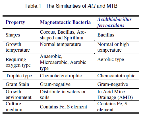
MTB can absorb the element of Fe from outer environment to synthesize intracellular nano-sized magnetosomes [14]. The growth environment of Acidthiobacillus ferrooxidans (At.f) contains abundant of the element of Fe and S that is the energy source of it. There are also many similarities between MTB and At.f (Table.1). Therefore,we suppose the growth environment of At.f is benefit to synthesize magnetosomes and At.f may contain some magnetotactic strain. We observed the magnetotactic of At.f and extracted magnetosomes from At.f. The results show that the At.f do contain magnetosomes. It is weak magnetotactic just because the existence of magnetosomes. We isolated the At.f with different magnetic field and research the effect of magnetic field act on At.f.
2. Materials and Methods
2.1 Materials
Strain: Isolated from AMD of Yunnan Dongchuan Mine.
9k culture medium: The concentrations of (NH4)2SO4,KCl,K2HPO4,MgSO4·7H2O,Ca(NO3)2,FeSO4·7H2O are: 3.00g/l,0.10g/l,0.50g/l,0.50g/l,0.01g/l,44.2g/l respectively.
Experimental apparatus: Galen III Microscope; SHT-III Tesla Meter; B-100 Automatic Ice Cube Maker; JY92-II- Ultrasonic Crasher; Eppendorf Centrifuge 5804R; WH-2 Micro Swirling Mixing Device.
2.2 Methods
2.2.1 Observation of the At.f’s magnetotactic property
The culture of At.f was collected by centrifugation and the iron ion was removed with H2SO4 of pH value 2.0. Then movement of the At.f was observed by using microscope under a magnetic field to determine whether At.f is magnetotactic.
2.2.2 Preparation of At.f cell’s suspending liquid
The culture that harvest at exponential phase is centrifugated by centrifuge for 5 minutes at 3000 rpm,then pour out the supernatant fluid,abandon the precipitate. The supernatant fluid is centrifugated for 10 minutes at 10000 rpm to collected At.f. Then pour out the supernatant fluid,added dilute sulfuric acid which the pH value is 2.0 to suspend the At.f,shaking for 5 minutes to dissolve Fe precipitate. Centrifugation,suspension and shaking procedure should repeat for about 10 times to clear the ion of iron out of the At.f cell completely.
2.2.3 Extraction of magnetosomes
Being suspended in the 10ml PBS buffer liquid,At.f was crashed (600 W/cm2,2.5 mins,30 pluses) by Ultrasonic crasher surrounded with ice. The specimen was alternated freeing and thawing for three times before crushing. Then we used magnetic iron close the bottom of beaker bottom to attract the magnetosomes When the cell wall had been broken up by ultrasonic crasher [15,16],the magnetosomes were cleaned by PBS buffer again and again until no ion of iron inside. It is important that the magnetic iron can't leave the bottom of beaker when pour the liquid. Then 0.5ml PBS buffer was added to suspend the magnetosomes (the raw specimen) in the beaker.
2.2.4 Detection of Fe ion in magnetosomes
We added a few 1mol/l HCl in PBS buffer which suspend the magnetosomes and shaked the beaker lightly to make the compound of iron dissolved. Then added two drops of 0.2% 0.5 ml KSCN in the beaker after the compound of iron dissolved completely with adding two drop of. If the solution’s color turned red,we concluded that the solution contained the ion of iron and At.f contained magnetosomes.
2.2.5 Purification of magnetosomes
The magnetosomes was extracted with the abovementioned method again,and was suspended it in the 0.5 ml PBS buffer. It was purified by recovering the magnet particle band after being centrifuged by 40%~80% sucrose gradient centrifugation (6000 rpm,10~20min) and being suspended in the PBS buffer. The magnetosomes suspended liquid was scattered by ultrasonic wave and washed by PBS buffer and ethanol to clean the cell fragment which absorbed on the surface of magnetosomes. These processes repeated 15 times to clean the magnetosomes completely and the magnetite was used to magnetize the magnetosomes by close up the bottom of beaker.
2.2.6 Extraction flowchart
The flowchart of magnetosomes’ extraction from At.f is shown in Figure 1.

Figure 1. The flowchart of extract magnetosomes
3. Results and Discussion
According to the present study,most of the MTB are heterotrophic bacteria and are difficult to culture because it needs anaerobic environment. But At.f are autotrophic,aerobic bacteria and are easy to culture. It is the first time we observe At.f is weak magnetotactic,extracted and detected the intracellular magnetosomes. The experiment results show us the followings:
a. The bacteria which extracellular iron ion was not removed were observed under the magnetic field function by using the microscope,we found the magnetotactic of At.f isn’t very obvious and majority’s movement is irregular. It probablely because that the substance contains Fe which blocks off the movement of At.f or that the material of the weak magnetism synthesized by cell weaken the external magnetic field to influence the cells’ movement. Most of the At.f cleaned by pH2.0 H2SO4 shows magnetotactic obviously. But there still has a few bacteria’s movement is irregular,it may because the number of intracellular magnetosomes is different.
b. The magnetosomes can be extracted from At.f. Just because it contains magnetosomes so that At.f is weak magnetotactic. We can use this magnetotactic to isolate At.f,we can also investigate the growth of At.f under the influence of mineral magnetism.
c. The magnetosome mainly contains Fe element. From Figure 2 we can see the magnetosomes can be aparted to monomer by sucrose density gradient centrifugation and ultrosonic scattering. The results show us that At.f do contain magnetosomes.

Figure 2. The electron microscope photo of magnetosomes. Bottoms are purified magnetosomes.
Acknowledgements
This work is supported by the Natural Science Foundation of China (NSFC) (No. 50374076).
References
- Blakemore R.P. (1975) Magnetotactic bacteria. Science,190: 377-379.
- Frankel R.B,Blakeinore R.P. and Wolfe R.S. (1979) Magnetite in freshwater magnetotactic bacterla. Science,203: 1355-1356.
- Blakemore R.P,Maratea D. and Wolfe R.S. (1979) Isolation and pure culture of a freshwater magnetic spirillum in chemically defined medium. J Bact,142: 720��729.
- Moench T.T. and Konetaka W.A. (1978) A novel method for isolation and study of a magnetotactic bacterium. Arch Microbiol,119: 203-212.
- Fassblnder J.W.E.,Stanjek H. and Vali H. (1990) Occurrence of magnetic bacteria in soil. Nature,343: 161-163.
- Matsunaga T.,Kawasaki M.,Xie Y.,et al. (1996) Chemiluminescence enzyme immunoassay using bacterial magnetic particles. Anal Chem,68(20): 3551-3554.
- Xie Y. (2003) Isolate,enrich and detect the carcinoembryonic antigen by using magnetotactic bacteria particle.Chin J Microbiol Immunol,23(2): 159-160.
- Fan G.C.,Li R.S.,et al. (1996) A research on distribution of magnetotactic bacteria and magnetosomes in China. Chinese Science Bulletin,41(4): 349-352.
- Gao J.,Xiao T.,et al. (2004) Isolation of a Novel Marine Magnetotactic Bacterium YSC-1 and Studies on the Highly Uniform,Magnetic Nanomaterial Magnetsome. High Technology Letters. 14(5): 44-47.
- Toshifumi Sakaguchi et al. (1996) A novel method for isolation of magnetic bacteria without magnetic collection using magnetotaxis. Journal of Microbiological Methods,26: 139-145.
- Blakemore R.P. (1982) Magnetotactic bacterla. Ann Rev Microbiol,36: 217-238.
- Blakemore R.P,Blakemore N.A.,Bazylinski D.A.,et al. (1989) Magnetotactic bacterla. In: Bergey’S Manual of Systematic Bacteriology,Murray R.G.E. ed. 3: 1882-1889.
- Matsunaga T.,Sakaguchi T. and Tadokoro F. (1991) Magnetite formation by a magnetic bacterium capable of growing aerobically. Appl Microbiol Biotech,35: 651-655.
- Tan H.,Feng D.W.,et al. (2000) Carrier Separation of Magnetic by Using Magnetotactic Bacteria. Journal of Foreign Ore Mineral Separation,(2): 2-5.
- Fu G.,Jiang W.,et al. (2004) Electron microscopic observation of magnetosomes formation in agnetospirillum gryphiswaldense and its purif ication. China Journal of Modern Medicine,14(5): 45-49.
- Ruan Y.,Tan Z.J.,et al. (1998) A Study of the Isolation of Magnetotactic Bacteria. Journal of Hunan Agricultural University,24(3): 238-240.


Open Access Journals
- Aquaculture & Veterinary Science
- Chemistry & Chemical Sciences
- Clinical Sciences
- Engineering
- General Science
- Genetics & Molecular Biology
- Health Care & Nursing
- Immunology & Microbiology
- Materials Science
- Mathematics & Physics
- Medical Sciences
- Neurology & Psychiatry
- Oncology & Cancer Science
- Pharmaceutical Sciences